Molecular visualization of synaptic fusion showing SNARE proteins and calcium ions.

Decoding the Secrets of Neurotransmitter Release: A New Look at Synaptic Fusion

"Cryo-electron tomography reveals the intricate dance of proteins during neurotransmitter release, offering fresh insights into this essential process."


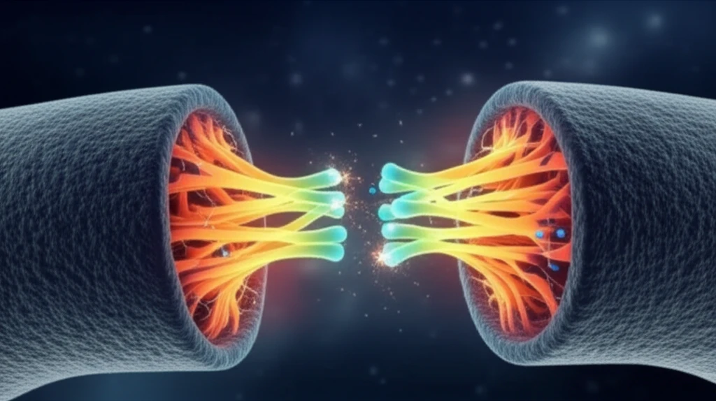
Neurotransmitter release, a fundamental process in neuronal communication, relies on the precise fusion of synaptic vesicles with the plasma membrane. This event, triggered by calcium ions, releases neurotransmitters into the synaptic cleft, enabling signal transmission between neurons. Understanding the molecular mechanisms underlying this fusion process is crucial for deciphering the complexities of brain function.

For years, scientists have been meticulously identifying and characterizing the essential components involved in neurotransmitter release. In vitro reconstitution experiments, simulating the fusion of two liposomes (one mimicking a synaptic vesicle, the other the plasma membrane), have provided valuable insights. Now, a groundbreaking study by Gipson et al. takes a leap forward by directly visualizing the steps in this fusion process using advanced electron cryotomography (cryo-ET).

Their research, published in PNAS, unveils that productive fusion occurs through point contacts between membranes, mediated by a small number of fusion-protein complexes—likely just two. This innovative approach offers a novel perspective on the intricate dance of proteins that orchestrate membrane fusion.

The Point of Contact: How Lipid Bilayers Fuse

Molecular visualization of synaptic fusion showing SNARE proteins and calcium ions.

Lipid bilayers, the building blocks of cell membranes, naturally repel each other through a “hydration force” when they come within a certain proximity (around 15-20 Å). Overcoming this repulsion is essential for membrane fusion, requiring a force that either acts in bulk or through localized fusion facilitators.

Computational studies suggest that creating a localized contact point minimizes the hydration force by reducing the area of closely apposed membranes. This reduction lowers the kinetic barrier and leads to the formation of a “hemifusion stalk,” a likely intermediate step in fusion. The cryo-ET images obtained by Gipson et al. support these theoretical predictions.

  • Key Players: SNAREs. The essential fusogens for synaptic vesicle fusion are SNARE proteins: synaptobrevin-2 (VAMP2) on the vesicle, syntaxin-1 on the plasma membrane, and SNAP-25 associated with the plasma membrane.
  • Calcium's Role. Synaptotagmin-1, a calcium sensor on the vesicle, initiates fusion when calcium is present.
  • Regulatory Factors. Complexin and Munc13 further modulate the fusion process, affecting the speed, yield, and range of morphological changes.
Cryo-ET, similar to medical tomography, reconstructs a 3D image from multiple projected views. However, electron damage limits exposure, making each image noisy and reducing achievable resolution. To improve signal-to-noise, researchers use phase-contrast imaging with Volta phase plates, enhancing the visibility of these pure phase objects. Gipson et al. describe “point contacts” – thin bridges of assembled protein fusion machinery – between liposomes bearing vesicle and plasma-membrane proteins, as well as “long contacts” with more extended density regions. The addition of regulatory proteins restricts the contacts to the former class, and calcium induces fusion on a millisecond timescale, causing point contacts to disappear.

Drawing Parallels: SNAREs Versus Viral Fusion

The point contacts observed by Gipson et al. are believed to represent the physiologically functional state, with complexin and Munc13 ensuring fidelity by modulating SNARE-complex geometry. The researchers estimate that just two complete, calcium-triggered complexes are sufficient for a productive fusion event.

Viral membrane fusion shares similarities with SNARE-mediated fusion but occurs on a longer timescale. Both processes involve a zippering-like rearrangement of fusion complexes, but unlike the preassembled SNARE complex, viral fusion proteins must come together after a triggering event. This difference accounts for the longer delay in viral fusion, highlighting the efficiency of preassembly in neurotransmitter release.

Cryo-ET images of influenza virus fusing with liposomes reveal intermediate structures that support this model. Visualizing the rapid transition from a calcium-triggered complex (or critical accumulation of bridging viral-protein intermediates) to a hemifusion stalk remains a formidable challenge for future research, promising deeper insights into the fundamental mechanisms of membrane fusion.

About this Article -

This article was crafted using a human-AI hybrid and collaborative approach. AI assisted our team with initial drafting, research insights, identifying key questions, and image generation. Our human editors guided topic selection, defined the angle, structured the content, ensured factual accuracy and relevance, refined the tone, and conducted thorough editing to deliver helpful, high-quality information.See our About page for more information.

This article is based on research published under:

DOI-LINK: 10.1073/pnas.1712038114, Alternate LINK

Title: Pictures Of The Prologue To Neurotransmitter Release

Subject: Multidisciplinary

Journal: Proceedings of the National Academy of Sciences

Publisher: Proceedings of the National Academy of Sciences

Authors: Stephen C. Harrison

Published: 2017-08-15

Everything You Need To Know

1

What is neurotransmitter release and why is it important?

Neurotransmitter release is a fundamental process in neuronal communication, where neurotransmitters are released into the synaptic cleft. This release, triggered by calcium ions, enables signal transmission between neurons. The research highlights the importance of understanding the molecular mechanisms behind this process for deciphering brain function. This is crucial for understanding how the brain works at a fundamental level, as it is the basis for how neurons communicate with each other.

2

What are the main components involved in synaptic vesicle fusion?

The key players in synaptic vesicle fusion are SNARE proteins: synaptobrevin-2 (VAMP2) on the vesicle, syntaxin-1 on the plasma membrane, and SNAP-25 associated with the plasma membrane. These proteins are essential fusogens, meaning they facilitate the fusion of the vesicle and plasma membranes. They are critical for the point contacts that are observed and the subsequent release of neurotransmitters. Without these proteins, the fusion process could not occur, and neuronal communication would be disrupted.

3

How is cryo-electron tomography used to study neurotransmitter release?

Cryo-electron tomography (cryo-ET) is an advanced imaging technique used to visualize the intricate steps in neurotransmitter release. It's like a 3D imaging method, similar to medical tomography, but uses electrons. This technique allows researchers to observe the point contacts and other protein interactions that facilitate membrane fusion. The advantage of cryo-ET is its ability to visualize the fusion process directly, providing insights into the molecular mechanisms at play. This is significant because it offers a novel perspective on how the dance of proteins orchestrates membrane fusion.

4

What role does calcium play in neurotransmitter release?

Calcium's role is as a trigger for synaptic vesicle fusion. Synaptotagmin-1, a calcium sensor on the vesicle, initiates fusion when calcium ions are present. This triggers the fusion process on a millisecond timescale. The presence of calcium is the signal that prompts the fusion machinery to work, allowing the release of neurotransmitters. Without calcium, the process would not initiate, and no signal transmission would happen.

5

What are point contacts, and what is their significance in the context of neurotransmitter release?

The point contacts are believed to represent the physiologically functional state, with complexin and Munc13 ensuring fidelity by modulating SNARE-complex geometry. The researchers estimate that just two complete, calcium-triggered complexes are sufficient for a productive fusion event. Complexin and Munc13 are regulatory factors that affect the speed, yield, and range of morphological changes during fusion. They work together to ensure the proper and efficient release of neurotransmitters. They also restrict the contacts to point contacts.

Newsletter Subscribe

Subscribe to get the latest articles and insights directly in your inbox.