High-tech temporal bone imaging scanner with glowing inner ear details

Decoding Temporal Bone Imaging: A Clearer Picture for Better Care

"Experts shed light on advancements in temporal bone imaging, offering new insights for diagnosing and treating related conditions."


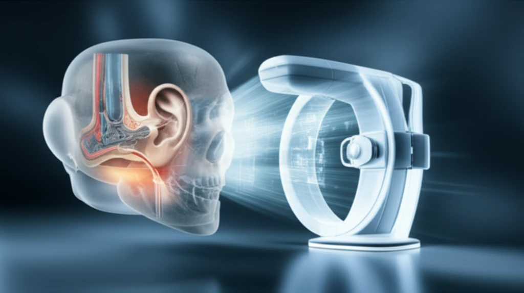
Imaging the temporal bone presents unique challenges for radiologists. Historically, conventional tomography was the standard, but advancements in computed tomography (CT) and magnetic resonance (MR) imaging have revolutionized the field, providing clearer and more detailed visualizations.

These modern imaging techniques allow for a more accurate assessment of the anatomy, overcoming the limitations of earlier methods. This evolution is critical because the temporal bone's complex structure houses vital components of the auditory and vestibular systems.

This article will discuss how temporal bone imaging has been enhanced by the contributions of experts in neuroimaging, offering insights into diagnosing and managing a range of conditions from congenital anomalies to traumatic injuries.

The Evolution of Temporal Bone Imaging

High-tech temporal bone imaging scanner with glowing inner ear details

The transition from conventional tomography to CT and MR imaging has significantly improved the diagnostic capabilities in temporal bone assessment. While earlier methods provided limited visualization, modern techniques offer detailed anatomical insights that were previously unattainable.

This enhanced visualization is crucial for accurately diagnosing a wide range of temporal bone conditions, including:

  • Congenital anomalies
  • Traumatic injuries
  • Infections
  • Neoplastic maladies
The incorporation of knowledge from otologic surgeons further refines interpretations, ensuring that imaging findings directly and positively impact patient care. Expertly authored articles contribute to a deeper understanding of the complexities involved.

The Future of Temporal Bone Imaging

Temporal bone imaging has drastically evolved, offering radiologists and surgeons improved diagnostic precision. The collaboration between radiologists and otologic surgeons enhances the clinical relevance of imaging interpretations, leading to better-informed treatment decisions.

Special thanks are extended to Amy and Gul for guest editing this important topic, their contributions and the work of other talented individuals are shaping the future of Head and Neck radiology. Their expertise ensures that advancements in imaging directly translate to improved patient outcomes.

Continued innovation and collaboration in this field promise even more refined diagnostic tools, ultimately benefiting patients and advancing the understanding of temporal bone disorders.

About this Article -

This article was crafted using a human-AI hybrid and collaborative approach. AI assisted our team with initial drafting, research insights, identifying key questions, and image generation. Our human editors guided topic selection, defined the angle, structured the content, ensured factual accuracy and relevance, refined the tone, and conducted thorough editing to deliver helpful, high-quality information.See our About page for more information.

Everything You Need To Know

1

What advancements have been made in temporal bone imaging?

Temporal bone imaging has evolved significantly, with modern techniques like Computed Tomography (CT) and Magnetic Resonance (MR) imaging replacing conventional tomography. These modern methods offer clearer, more detailed visualizations, leading to more accurate diagnoses. CT and MR imaging surpass the limitations of earlier methods, providing crucial insights into the temporal bone's complex structures. This enhanced imaging allows for a more precise assessment of anatomy, directly impacting patient care by enabling better detection of conditions.

2

Why is the evolution of temporal bone imaging important?

The transition from conventional tomography to CT and MR imaging is important because the temporal bone houses the auditory and vestibular systems. CT and MR imaging provide detailed anatomical insights which is crucial for accurate diagnosis and management of conditions affecting these systems. This leads to improved diagnostic accuracy and patient care in otology and neuro-otology. The improved visualization allows for the detection of congenital anomalies, traumatic injuries, infections, and neoplastic maladies.

3

What were the limitations of conventional tomography?

The limitations of conventional tomography include providing limited visualization of the temporal bone's intricate structures. This hindered accurate diagnosis and treatment planning. Earlier methods did not offer the detailed anatomical insights provided by CT and MR imaging, which are essential for assessing conditions affecting the auditory and vestibular systems. The switch to modern techniques allows radiologists to see more, leading to better patient outcomes.

4

Why is collaboration between radiologists and otologic surgeons important?

The collaboration between radiologists and otologic surgeons is vital because it refines the interpretation of imaging findings. This collaboration ensures that the imaging results are clinically relevant and directly impact patient care. Expert input from otologic surgeons helps translate imaging findings into actionable treatment plans, leading to better-informed decisions and improved patient outcomes.

5

What types of conditions are diagnosed using temporal bone imaging?

Temporal bone imaging is crucial for diagnosing and treating various conditions, including congenital anomalies, traumatic injuries, infections, and neoplastic maladies. These conditions affect the delicate structures within the temporal bone, housing the auditory and vestibular systems. Accurate imaging allows for early detection, precise diagnosis, and targeted treatment, ultimately improving patient outcomes by enhancing the management of these conditions.

Newsletter Subscribe

Subscribe to get the latest articles and insights directly in your inbox.