Decoding Spinal Cord Injury: How MRI Can Predict Recovery
"New research unveils how MRI technology can assess white matter damage after spinal cord injury, offering insights into recovery potential and treatment strategies."
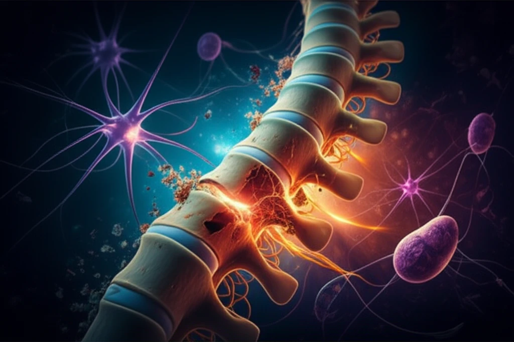
Spinal cord injuries (SCI) present a significant challenge, often leading to sensory and motor dysfunction, impacting the lives of many. The damage that occurs after an SCI unfolds in two phases: the primary phase, where initial trauma inflicts immediate harm, and the secondary phase, characterized by edema, inflammation, and tissue degeneration. These secondary injuries can exacerbate the initial damage, particularly affecting the white matter, which is crucial for transmitting signals throughout the nervous system.
Understanding the extent of white matter damage is key to predicting recovery after an SCI. White matter contains bundles of nerve fibers (axons) coated with myelin, which speeds up signal transmission. When this area is damaged, it disrupts communication between the brain and body. Scientists are actively seeking ways to assess this damage non-invasively, allowing for more accurate prognoses and targeted treatments.
Diffusion Tensor Imaging (DTI), a specialized type of MRI, has emerged as a promising tool for evaluating white matter integrity. Unlike conventional MRI, DTI can reveal subtle microstructural changes in the white matter by tracking the movement of water molecules within the tissue. This article explores recent research that uses DTI to investigate white matter damage in rats with severe spinal cord injuries, demonstrating its potential to predict functional recovery.
MRI's Role in Assessing White Matter Post-SCI
Researchers at Capital Medical University used a 7.0T MRI scanner to image the spinal cords of rats before and after inducing a severe contusion SCI. They focused on specific regions of interest (ROIs) within the white matter, including the dorsal column (DC), left and right lateral white matter (LLWM and RLWM), and left and right ventral white matter (LVWM and RVWM). By analyzing DTI parameters such as fractional anisotropy (FA), mean diffusivity (MD), axial diffusivity (AD), and radial diffusivity (RD), they were able to quantify the extent of white matter damage in each region.
- Fractional Anisotropy (FA): Measures the directionality of water diffusion. Lower FA values indicate more disrupted white matter.
- Mean Diffusivity (MD): Reflects the overall magnitude of water diffusion. Higher MD values can indicate edema or tissue damage.
- Axial Diffusivity (AD): Measures water diffusion parallel to the nerve fibers. Reduced AD suggests axonal damage.
- Radial Diffusivity (RD): Measures water diffusion perpendicular to the nerve fibers. Increased RD can indicate myelin damage.
Implications for Future SCI Treatment
The findings suggest a critical "window of opportunity" in the first three days after SCI for intervention. Protecting and preserving the spared white matter during this period may significantly improve long-term motor function recovery. The ventral column, in particular, appears to play a crucial role.
Furthermore, this research supports DTI as a valuable, non-invasive tool for diagnosing the severity of acute SCI and predicting functional outcomes. This could enable clinicians to tailor treatment strategies based on individual patient profiles.
While this study was conducted in rats, the implications for human SCI are significant. By using DTI to assess white matter damage, clinicians may be able to develop more effective and personalized rehabilitation plans, ultimately improving the lives of individuals affected by SCI.