Ultrasound image overlayed on a ram, showcasing internal structures.

Decoding Sheep Fertility: The Role of Ultrasound in Young Ram Health

"A non-invasive approach to monitoring reproductive development and ensuring flock productivity."


In the dynamic world of livestock management, enhancing reproductive efficiency stands as a cornerstone of boosting overall productivity. Breeders are constantly seeking innovative methods to refine their practices and maximize flock potential. Among the array of emerging biotechnologies, ultrasonography is proving to be a game-changer in veterinary reproductive health.

Ultrasound imaging, or US, offers a simple yet powerful means of complementing traditional clinical reproductive exams. By providing detailed visualizations of internal structures, it allows for a more comprehensive assessment of reproductive health, leading to increased accuracy in diagnoses and more effective management strategies. This non-invasive technique is paving the way for a new era of precision in animal husbandry.

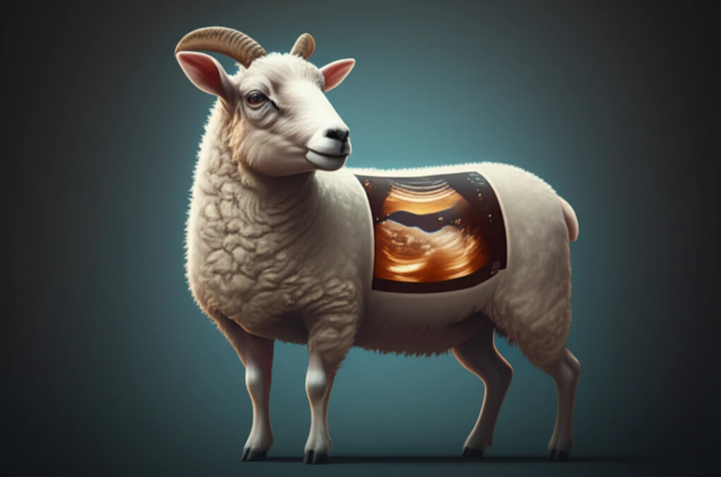
While ultrasonography has become a standard tool in andrology, its application in ovine (sheep) reproductive health remains relatively unexplored. Specifically, the normal ultrasonographic characteristics of young, developing rams are not well-documented, making it challenging to establish benchmarks for assessing reproductive potential. This article delves into a groundbreaking study that aims to address this gap by describing the ultrasonographic findings in the testicles and epididymides of clinically healthy, young hair sheep.

What Can Ultrasound Reveal About Ram Fertility?

Ultrasound image overlayed on a ram, showcasing internal structures.

A study published in the Arq. Bras. Med. Vet. Zootec. journal investigated the ultrasonographic features of testicles and epididymides in young, healthy hair sheep. The study tracked the development of 18 crossbred lambs (Dorper x Santa Inês) from 140 to 280 days of age. Over this period, researchers conducted biometrical measurements of the testicles and performed regular ultrasound examinations to monitor their development.

The researchers used a Pie Medical Falco 100 ultrasound machine with an 8.0 MHz linear array transducer to visualize and document the testicular structures. They carefully evaluated the echotexture of the testicular parenchyma and mediastinum, as well as the epididymis and pampiniform plexus. These measurements were then correlated with the animals' age and reproductive development.

Key findings from the study include:
  • Testicular Parenchyma: The testicular parenchyma displayed a homogeneous echogenicity (low to moderate), which increased with age.
  • Mediastinum Testis: The echogenicity and thickness of the mediastinum testis also increased with age.
  • Epididymis: The tail of the epididymis appeared hypoechoic compared to the testicular parenchyma.
  • Calcifications: Mild calcifications were observed in the testicular parenchyma of five lambs.
These findings offer valuable insights into the normal ultrasonographic appearance of the testicles and epididymides in young rams. By establishing these baseline characteristics, veterinarians and breeders can more effectively use ultrasound to monitor testicular health, detect abnormalities, and predict reproductive potential.

Why Ultrasound Matters for Sheep Breeders

The study concludes that ultrasonography is a valuable tool for monitoring the morphophysiological characteristics of the external genitalia in sheep. It enables the identification and tracking of progressive physiological changes in the testicles and epididymides of young, clinically healthy woolless rams. This ultimately contributes to better management of flock fertility and overall reproductive success.

About this Article -

This article was crafted using a human-AI hybrid and collaborative approach. AI assisted our team with initial drafting, research insights, identifying key questions, and image generation. Our human editors guided topic selection, defined the angle, structured the content, ensured factual accuracy and relevance, refined the tone, and conducted thorough editing to deliver helpful, high-quality information.See our About page for more information.

Everything You Need To Know

1

How does ultrasound improve sheep breeding practices compared to traditional methods?

Ultrasound technology provides a non-invasive method for visualizing internal structures, offering a more comprehensive assessment of reproductive health in rams. Traditional methods often rely on external examinations, which may not reveal subtle abnormalities. By using ultrasound, breeders gain detailed insights into the testicles and epididymides, allowing for increased accuracy in diagnoses and more effective management strategies. This leads to better predictions of reproductive potential and overall flock fertility, exceeding the limitations of traditional approaches.

2

What specific testicular structures do researchers evaluate using ultrasound in young rams?

The study focuses on evaluating the ultrasonographic features of the testicular parenchyma, mediastinum testis, and epididymis. The testicular parenchyma's echogenicity (how it reflects ultrasound waves) and the mediastinum testis's echogenicity and thickness are carefully examined. The tail of the epididymis is also assessed. These assessments are correlated with the ram's age and reproductive development, providing insights into the health and maturity of the reproductive system.

3

What are the key ultrasonographic findings in young, healthy hair sheep, and how do they change with age?

The key findings include the following. First, the testicular parenchyma displays a homogeneous echogenicity that increases with age. Second, the echogenicity and thickness of the mediastinum testis also increase with age. Third, the tail of the epididymis appears hypoechoic compared to the testicular parenchyma. Finally, mild calcifications were observed in the testicular parenchyma of some lambs. These findings establish benchmarks for assessing testicular health and predicting reproductive potential as the rams mature.

4

How can ultrasound help in detecting abnormalities in young rams, and what are the implications?

Ultrasound allows veterinarians and breeders to monitor the morphophysiological characteristics of the external genitalia in sheep. It enables the identification of abnormalities, such as variations in echogenicity or structural irregularities in the testicles and epididymides. Early detection is crucial because it can predict reproductive potential. Identifying issues early enables breeders to make informed decisions about breeding strategies, potentially improving flock fertility and overall reproductive success.

5

Beyond identifying physical structures, what additional data does ultrasound provide that benefits sheep breeders?

Besides visualizing the structures, ultrasound measurements are correlated with the rams' age and overall reproductive development. This means breeders can track progressive physiological changes in the testicles and epididymides over time. This data helps establish baselines for normal development and identify deviations that may impact fertility. Furthermore, by understanding these changes, breeders can better manage their flocks, improve breeding efficiency, and ultimately enhance overall productivity and profitability.

Newsletter Subscribe

Subscribe to get the latest articles and insights directly in your inbox.