AI analyzing microscopic muscle fibers

Decoding Muscle Health: How AI is Revolutionizing Muscular Dystrophy Analysis

"A breakthrough neural network approach promises faster, more accurate diagnosis and monitoring of muscle diseases."


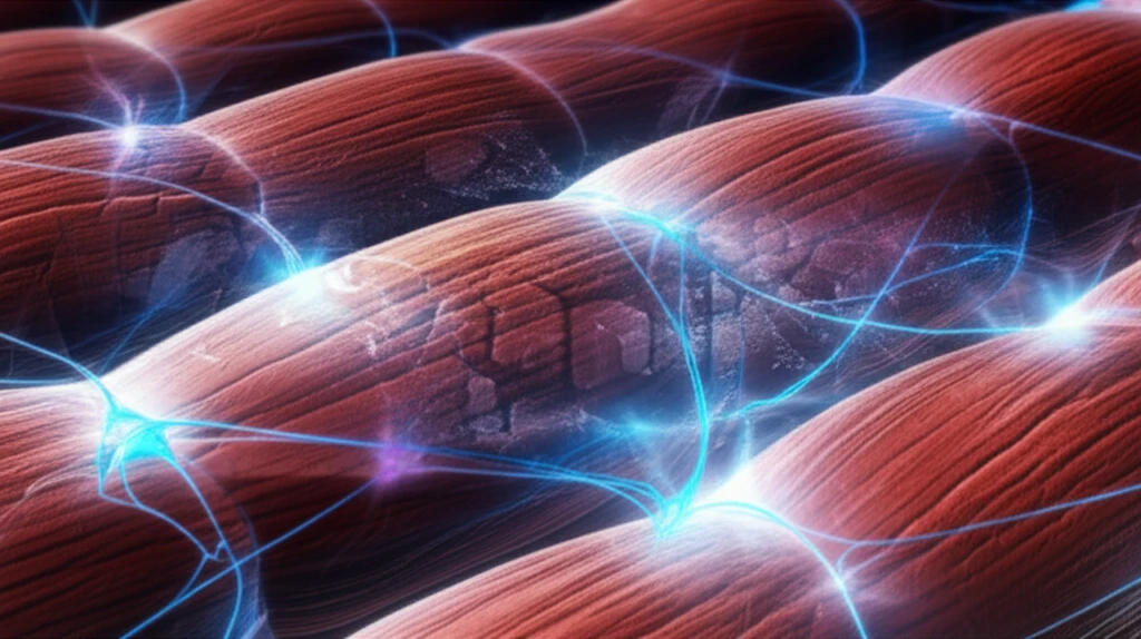
Muscular dystrophies, a group of over 40 diseases, significantly impair skeletal muscle function. A key characteristic is often the deficiency of dystrophin, a protein vital for muscle fiber integrity. This deficiency leads to porous membranes and eventual loss of cytoplasm, directly impacting muscle health and regenerative capacity. Analyzing the morphology of muscle fiber cross-sections is critical for assessing the severity and progression of these diseases.

However, traditional manual analysis of muscle fiber images is labor-intensive and prone to variability between observers. The sheer number of objects and the subtle differences in their morphology make it challenging to obtain consistent and reliable results. This highlights the need for automated solutions that can provide objective and reproducible analysis.

Enter artificial intelligence. Researchers have developed a deep convolutional neural network (DCNN) approach, enhanced by post-processing techniques, to detect and measure muscle fiber cross-sections. This innovative method offers a promising alternative for analyzing histopathological images, particularly those with low signal-to-noise ratio, uneven backgrounds, and closely spaced muscle fibers—conditions commonly found in diseased tissue.

AI-Powered Muscle Fiber Analysis: How Does It Work?

AI analyzing microscopic muscle fibers

The AI system employs a sophisticated DCNN architecture, drawing upon the strengths of networks like U-net and FusionNet. These networks are trained to segment muscle fiber boundaries in microscopic images. The process involves:

  • Image Enhancement: Correcting defects and inconsistencies in staining quality to ensure clear visibility of muscle fibers.

  • Seed Detection: Identifying the centers of muscle fibers to guide boundary segmentation.
  • Boundary Segmentation: Delineating the precise borders of each muscle fiber using advanced algorithms.
The AI network then analyzes these segmented images to extract key morphological characteristics, such as cross-sectional area, shape, and fiber density. This data provides valuable insights into the health and regenerative capacity of the muscle tissue.

The Future of Muscle Disease Diagnosis

This AI-driven approach holds significant promise for improving the diagnosis and monitoring of muscular dystrophies and other muscle-related conditions. By providing faster, more objective, and more reproducible analysis, it can empower researchers and clinicians to better understand these diseases and develop more effective treatments.

About this Article -

This article was crafted using a human-AI hybrid and collaborative approach. AI assisted our team with initial drafting, research insights, identifying key questions, and image generation. Our human editors guided topic selection, defined the angle, structured the content, ensured factual accuracy and relevance, refined the tone, and conducted thorough editing to deliver helpful, high-quality information.See our About page for more information.

This article is based on research published under:

DOI-LINK: 10.1016/j.compbiomed.2018.11.007, Alternate LINK

Title: A Neural Network Approach To Analyze Cross-Sections Of Muscle Fibers In Pathological Images

Subject: Health Informatics

Journal: Computers in Biology and Medicine

Publisher: Elsevier BV

Authors: Ye Li, Zhong Yang, Yaming Wang, Xinhua Cao, Xiaoyin Xu

Published: 2019-01-01

Everything You Need To Know

1

What are muscular dystrophies, and why is AI significant in their analysis?

Muscular dystrophies are a group of over 40 diseases that impair skeletal muscle function. A key characteristic is often the deficiency of dystrophin, a protein vital for muscle fiber integrity. This deficiency leads to porous membranes and eventual loss of cytoplasm, directly impacting muscle health and regenerative capacity. AI is important for understanding and treating these conditions because manual analysis is labor-intensive and prone to variability, hindering consistent and reliable results. This AI-driven approach provides faster, more objective, and reproducible analysis, empowering researchers and clinicians to better understand these diseases.

2

How does the AI system work in analyzing muscle fiber images?

The AI system uses a deep convolutional neural network (DCNN) architecture, drawing upon the strengths of networks like U-net and FusionNet. The process involves image enhancement to correct defects, seed detection to identify fiber centers, and boundary segmentation to delineate fiber borders. The AI network then extracts morphological characteristics like cross-sectional area, shape, and fiber density from the segmented images. The use of AI allows for the ability to analyze images with low signal-to-noise ratios, uneven backgrounds, and closely spaced muscle fibers.

3

What is the role of dystrophin in muscle health, and why is analyzing muscle fiber cross-sections important?

Dystrophin is a crucial protein for muscle fiber integrity. Its deficiency leads to porous membranes and loss of cytoplasm, which directly impacts muscle health and reduces regenerative capacity. The inability of the muscle to repair and maintain itself leads to the functional impairment characteristic of muscular dystrophies. Analyzing muscle fiber cross-sections helps assess the severity of the diseases because it reveals the extent of muscle damage and the progression of the disease.

4

What is a DCNN, and how is it used to analyze muscle fibers?

A DCNN is a sophisticated AI architecture that is trained to segment muscle fiber boundaries in microscopic images. This involves correcting staining defects (image enhancement), identifying fiber centers (seed detection), and delineating fiber borders (boundary segmentation). The AI then analyzes the segmented images to measure morphological characteristics. This process allows for detailed analysis of muscle tissue, providing critical information on the health and characteristics of the muscle fibers.

5

How does the AI-driven approach improve the diagnosis and monitoring of muscle diseases?

AI-powered analysis significantly improves the diagnosis and monitoring of muscle diseases by offering a faster, more objective, and reproducible method compared to traditional manual analysis. This automated approach can help researchers and clinicians to gain a better understanding of the disease progression and potentially enable the development of more effective treatments. The ability to analyze large datasets consistently is a major advantage, leading to more reliable conclusions and potentially accelerating the pace of research in the field.

Newsletter Subscribe

Subscribe to get the latest articles and insights directly in your inbox.