Surreal illustration of lung anatomy with algorithms for lung cancer treatment.

Decoding Lung Cancer: How Advanced Imaging and Algorithms Are Changing Treatment

"New research reveals how different imaging techniques and segmentation algorithms impact the accuracy and effectiveness of lung cancer diagnosis and treatment planning, offering hope for more personalized and effective care."


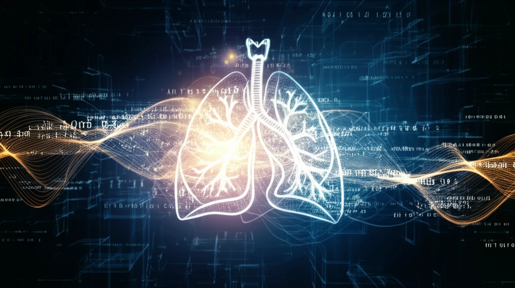
Lung cancer remains a significant global health challenge, necessitating continuous advancements in diagnostic and treatment strategies. Traditional methods often struggle with accurately delineating tumor boundaries and understanding the complex characteristics of cancerous tissues. However, with innovations in medical imaging and computational analysis, there's new hope for enhancing precision and personalization in lung cancer care.

The integration of advanced imaging technologies like 18F-FDG PET/CT (18-fluoro-2-deoxyglucose positron emission tomography/computed tomography) has revolutionized the ability to visualize and assess tumors. These scans provide crucial information about tumor heterogeneity, which is increasingly recognized as a key factor in predicting how a cancer will respond to treatment.

Recent research has focused on how different image segmentation algorithms affect the measurement and interpretation of PET/CT scans. These algorithms are vital for accurately outlining tumors, but the variability in their performance can impact the reliability of derived parameters used for diagnosis and prognosis. A study published in EJNMMI Research has shed light on these critical differences, offering insights that could refine clinical practices and improve patient outcomes.

The Impact of Segmentation Algorithms on Lung Cancer Diagnosis

Surreal illustration of lung anatomy with algorithms for lung cancer treatment.

The study published in EJNMMI Research investigated the effects of three different segmentation algorithms—freehand (FH), 40% of maximum intensity threshold (40P), and fuzzy locally adaptive Bayesian (FLAB)—on the measurement of texture parameters in non-small cell lung cancer (NSCLC) 18F-FDG PET/CT images. The goal was to compare these algorithms in terms of inter-observer reproducibility and prognostic capability. Fifty-three NSCLC patients were involved, and their scans were segmented by three expert readers using each of the algorithms.

Key findings from the study revealed significant differences in the reproducibility of the algorithms. The 40P algorithm demonstrated the highest inter-observer reproducibility, indicating more consistent results across different observers. This is crucial because consistent tumor delineation ensures that subsequent measurements and analyses are reliable, which directly impacts treatment planning and outcome prediction. In contrast, the FH and FLAB algorithms showed more variability, potentially leading to less reliable results in clinical settings.

Here are the highlights of the main differences observed in the study:
Understanding the nuances of these algorithms helps healthcare providers make informed decisions about which methods to use for specific cases. The study suggests that while different algorithms may delineate tumor volumes differently, their impact on survival models remains equivalent. This means that a 40% threshold algorithm can be a reliable choice for texture analysis of 18F-FDG PET in NSCLC, offering a balance between accuracy and reproducibility.

Personalized Treatment on the Horizon

The insights from this research underscore the importance of selecting appropriate imaging and segmentation techniques to ensure reliable and reproducible results. As the field of radiomics continues to evolve, standardized methodologies will be essential for translating research findings into clinical practice. By optimizing these processes, healthcare providers can better leverage the power of advanced imaging to deliver personalized and effective lung cancer care, ultimately improving patient outcomes and quality of life. Continuous research and collaboration will pave the way for more precise and tailored approaches to combat this challenging disease.

About this Article -

This article was crafted using a human-AI hybrid and collaborative approach. AI assisted our team with initial drafting, research insights, identifying key questions, and image generation. Our human editors guided topic selection, defined the angle, structured the content, ensured factual accuracy and relevance, refined the tone, and conducted thorough editing to deliver helpful, high-quality information.See our About page for more information.

This article is based on research published under:

DOI-LINK: 10.1186/s13550-017-0310-3, Alternate LINK

Title: The Effects Of Segmentation Algorithms On The Measurement Of 18F-Fdg Pet Texture Parameters In Non-Small Cell Lung Cancer

Subject: Radiology, Nuclear Medicine and imaging

Journal: EJNMMI Research

Publisher: Springer Science and Business Media LLC

Authors: Usman Bashir, Gurdip Azad, Muhammad Musib Siddique, Saana Dhillon, Nikheel Patel, Paul Bassett, David Landau, Vicky Goh, Gary Cook

Published: 2017-07-26

Everything You Need To Know

1

How are imaging techniques like 18F-FDG PET/CT scans revolutionizing lung cancer diagnosis and treatment?

Recent advancements integrate imaging technologies like 18F-FDG PET/CT to visualize and assess tumors, providing crucial information about tumor heterogeneity. This is important because tumor heterogeneity is a key factor in predicting how a cancer will respond to treatment. The research focuses on how different image segmentation algorithms affect the measurement and interpretation of PET/CT scans, which is vital for accurately outlining tumors. Variability in algorithm performance can affect the reliability of derived parameters used for diagnosis and prognosis. Therefore, selecting the appropriate imaging and segmentation techniques is essential for reliable and reproducible results.

2

What segmentation algorithms were compared in the EJNMMI Research study, and how did they impact the measurement of texture parameters in NSCLC patients?

The study published in EJNMMI Research evaluated three segmentation algorithms: freehand (FH), 40% of maximum intensity threshold (40P), and fuzzy locally adaptive Bayesian (FLAB). These algorithms were assessed for inter-observer reproducibility and prognostic capability using 18F-FDG PET/CT images from fifty-three NSCLC patients. The key findings highlighted significant differences in the reproducibility of these algorithms, which impacts the reliability of tumor delineation and subsequent measurements.

3

What were the key differences in inter-observer reproducibility among the freehand (FH), 40% of maximum intensity threshold (40P), and fuzzy locally adaptive Bayesian (FLAB) algorithms?

The 40P algorithm exhibited the highest inter-observer reproducibility, ensuring consistent results across different observers. This consistency is critical because it ensures that tumor delineation is reliable, which directly impacts treatment planning and outcome prediction. In contrast, the FH and FLAB algorithms showed more variability, which can lead to less reliable results in clinical settings. While the study indicated that the impact on survival models remained equivalent across algorithms, the reproducibility of 40P makes it a reliable choice for texture analysis of 18F-FDG PET in NSCLC.

4

How do insights from research on imaging and segmentation techniques contribute to personalized treatment strategies for lung cancer patients?

By optimizing imaging and segmentation processes, healthcare providers can leverage advanced imaging to deliver personalized and effective lung cancer care. The study indicates that methodologies will be essential for translating research findings into clinical practice. Continuous research and collaboration will pave the way for more precise and tailored approaches to combat this challenging disease, ultimately improving patient outcomes and quality of life.

5

How does radiomics factor into advanced imaging for lung cancer, and what role does it play in clinical decision-making?

Radiomics is the process of converting medical images into mineable data, to build models that assist clinical decision-making. While not detailed in the text, radiomics is connected because the research highlighted in the text underscores the importance of selecting appropriate imaging and segmentation techniques to ensure reliable and reproducible results. As the field of radiomics continues to evolve, standardized methodologies will be essential for translating research findings into clinical practice. Optimization of these processes allows healthcare providers to better leverage the power of advanced imaging to deliver personalized and effective lung cancer care, ultimately improving patient outcomes and quality of life.

Newsletter Subscribe

Subscribe to get the latest articles and insights directly in your inbox.