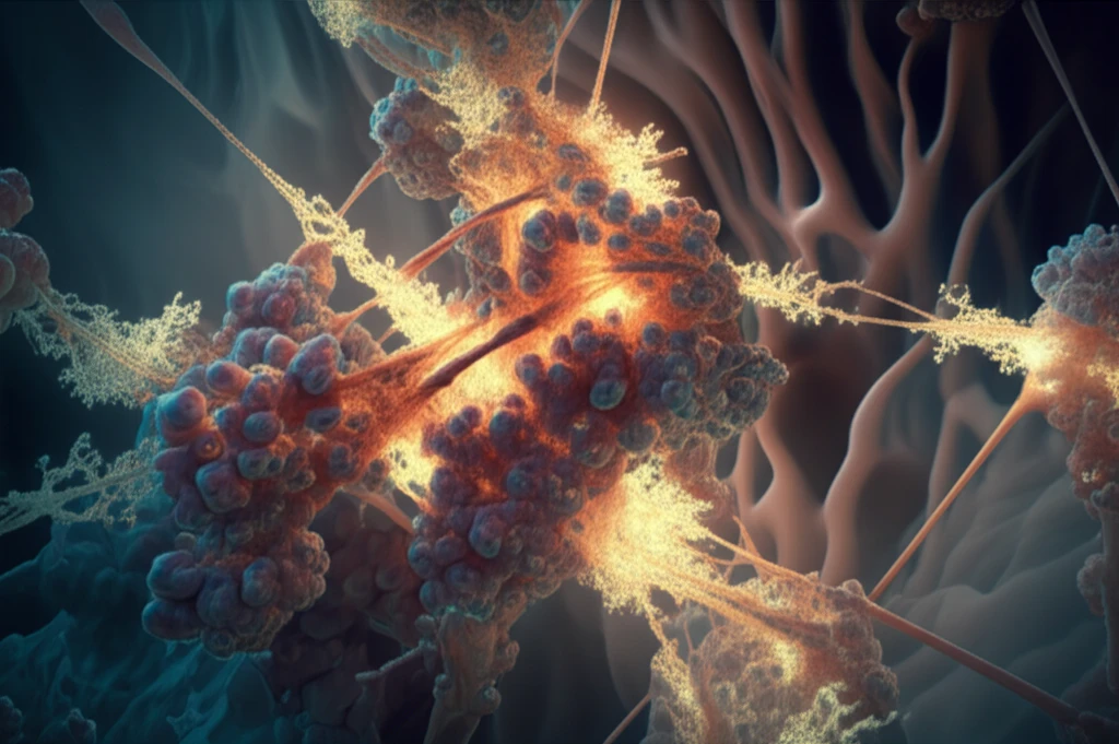
Microscopic calcium oxalate crystals intertwined with glowing, phosphorylated osteopontin peptides.

Decoding Kidney Stones: How Osteopontin Peptides and Phosphorylation Hold the Key

"A quantitative study reveals the crucial role of osteopontin peptide phosphorylation in kidney stone formation, offering new insights into prevention and treatment."


Kidney stones are a common and painful condition affecting millions worldwide. The primary mineral component of most kidney stones is calcium oxalate monohydrate (COM). Understanding how COM crystals form and aggregate is crucial in developing effective prevention and treatment strategies.

Recent research has focused on the role of polyelectrolyte-crystal interactions in biomineralization, which includes the formation of kidney stones. One key player in this process is osteopontin (OPN), a protein known to inhibit COM formation. However, the exact mechanisms by which OPN and its various forms influence kidney stone development are not fully understood.

A new quantitative study investigates how different phosphorylated forms of an osteopontin peptide interact with COM crystals. By examining the adsorption and incorporation of these peptides, the researchers aim to shed light on the role of phosphorylation in regulating kidney stone formation.

The Science Behind Kidney Stone Formation: Unveiling Osteopontin's Role

Microscopic calcium oxalate crystals intertwined with glowing, phosphorylated osteopontin peptides.

The study focuses on synthetic peptides corresponding to the amino acid sequence 220–235 of rat bone osteopontin. These peptides were created with varying degrees of phosphorylation: no phosphates (P0), one phosphate (P1), and three phosphates (P3). The researchers then observed how these peptides interacted with COM crystals formed in a controlled laboratory setting.

Quantitative fluorimetry was used to measure the adsorption and incorporation of the peptides into the COM crystals. Additionally, confocal and scanning electron microscopy provided detailed visual information about the crystal structures and peptide distribution. X-ray and Raman spectroscopy were employed to analyze the crystal composition and structure at a molecular level.

The key findings of the study revealed several important insights:
  • Higher phosphorylation leads to stronger binding: Peptides with more phosphate groups (P3) exhibited stronger and more irreversible adsorption to COM crystals compared to those with fewer phosphates (P1).
  • Increased incorporation with phosphorylation: P3 peptides were incorporated into the crystals at a significantly higher rate than P1 peptides, suggesting that phosphorylation promotes incorporation.
  • Face-specific interactions: Both adsorption and incorporation occurred preferentially on specific crystal faces, with the {100} face showing the strongest interaction.
Interestingly, high levels of P3 incorporation resulted in crystal cleavage, indicating that excessive incorporation of highly phosphorylated peptides can disrupt crystal structure. However, extrapolation of data from intact crystals showed that even in these cases, a substantial amount of P3 could be incorporated without causing structural damage. Spectroscopic analysis revealed that the incorporation of peptides did not significantly alter the overall crystal structure, suggesting that the peptides are surrounded by the growing crystal matrix and then incorporated.

Implications for Kidney Stone Treatment and Prevention

This study provides valuable insights into the complex interplay between osteopontin, phosphorylation, and calcium oxalate crystal formation. Understanding how different forms of OPN interact with COM crystals can pave the way for developing targeted therapies to prevent or dissolve kidney stones. By manipulating the degree of phosphorylation or using specific OPN peptides, it may be possible to control crystal growth and aggregation, ultimately reducing the risk of kidney stone formation. Further research is needed to explore these possibilities and translate these findings into clinical applications.

About this Article -

This article was crafted using a human-AI hybrid and collaborative approach. AI assisted our team with initial drafting, research insights, identifying key questions, and image generation. Our human editors guided topic selection, defined the angle, structured the content, ensured factual accuracy and relevance, refined the tone, and conducted thorough editing to deliver helpful, high-quality information.See our About page for more information.

This article is based on research published under:

DOI-LINK: 10.1007/s00240-018-01105-x, Alternate LINK

Title: Incorporation Of Osteopontin Peptide Into Kidney Stone-Related Calcium Oxalate Monohydrate Crystals: A Quantitative Study

Subject: Urology

Journal: Urolithiasis

Publisher: Springer Science and Business Media LLC

Authors: Jared S. Gleberzon, Yinyin Liao, Silvia Mittler, Harvey A. Goldberg, Bernd Grohe

Published: 2018-12-19

Everything You Need To Know

1

What are kidney stones made of?

Kidney stones are primarily composed of calcium oxalate monohydrate (COM) crystals. Understanding how these crystals form and aggregate is crucial for developing effective prevention and treatment strategies. The formation process involves interactions between COM crystals and molecules like osteopontin (OPN).

2

What is the role of osteopontin in the context of kidney stone formation?

Osteopontin (OPN) is a protein that plays a vital role in the formation of kidney stones. It is known for its ability to interact with calcium oxalate monohydrate (COM) crystals. The study investigates how different phosphorylated forms of an osteopontin peptide affect COM crystal formation. The key finding suggests that phosphorylation of OPN peptides influences their interaction with COM crystals, with higher phosphorylation leading to stronger binding and incorporation into the crystals.

3

What is phosphorylation and how does it relate to the study?

Phosphorylation refers to the addition of phosphate groups to a molecule, in this case, the osteopontin (OPN) peptides. The study examined peptides with varying degrees of phosphorylation: P0 (no phosphates), P1 (one phosphate), and P3 (three phosphates). The level of phosphorylation influences how the peptides interact with calcium oxalate monohydrate (COM) crystals. Higher phosphorylation (P3) was found to enhance binding and incorporation into the crystals.

4

Why is osteopontin peptide phosphorylation important?

The significance of osteopontin peptide phosphorylation lies in its potential for influencing the growth of calcium oxalate monohydrate (COM) crystals, which are the main components of kidney stones. Understanding how phosphorylation affects the interaction of osteopontin peptides with COM crystals can lead to the development of targeted therapies. The study's results suggest that manipulating phosphorylation levels could be a strategy to control crystal growth and aggregation, potentially reducing the risk of kidney stone formation.

5

What are the implications of this research for kidney stone treatment and prevention?

The implications of these findings are significant for the development of kidney stone treatments and preventative measures. The study indicates that the degree of osteopontin peptide phosphorylation affects the interaction with calcium oxalate monohydrate (COM) crystals. By understanding these mechanisms, it may be possible to design therapies that manipulate phosphorylation or utilize specific osteopontin peptides to control crystal growth. This could lead to methods for preventing or dissolving kidney stones, improving the lives of those affected by this painful condition.

Newsletter Subscribe

Subscribe to get the latest articles and insights directly in your inbox.