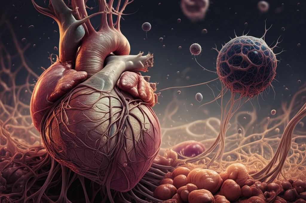
Surreal illustration of a heart with cytoskeletal network representing cardiac fibrosis.

Decoding Heart Failure: Can We Stop Fibrosis in Its Tracks?

"New research illuminates the role of cellular mechanics in hypofibrotic cardiac fibroblasts, offering potential pathways for treatment."


Heart failure is a complex condition where the heart can't pump enough blood to meet the body's needs. Often, this involves changes in the heart's structure, a process called remodeling. Hemodynamic load, the forces acting on the heart, plays a big role in this remodeling. There are two main types of hemodynamic load: pressure overload (increased afterload), often caused by high blood pressure, and volume overload (increased preload), where the heart has to pump more blood than usual.

While pressure overload is known to cause the heart muscle to thicken and become stiff, volume overload leads to a different kind of remodeling. The heart chambers enlarge, and the amount of supportive tissue, called the extracellular matrix, decreases. Cardiac fibroblasts (CFs) are the cells responsible for maintaining this matrix. Understanding how these cells behave under volume overload is crucial for developing effective treatments for heart failure.

New research sheds light on the behavior of cardiac fibroblasts in volume overload, revealing a unique 'hypofibrotic' phenotype. This means the cells produce less of the proteins that make up the extracellular matrix, leading to a weaker, more dilated heart. The study dives deep into the cellular mechanisms behind this phenomenon, focusing on the role of the cytoskeleton and potential therapeutic targets.

What's the Link Between Volume Overload and Cardiac Fibroblast Behavior?

Surreal illustration of a heart with cytoskeletal network representing cardiac fibrosis.

To investigate this, researchers created a model of volume overload in rats by creating an aortocaval fistula (ACF), a connection between the aorta and vena cava that increases blood volume returning to the heart. After four weeks, they isolated cardiac fibroblasts from these rats and compared them to cells from healthy control rats. The results were striking: the fibroblasts from volume overload hearts displayed a distinct hypofibrotic phenotype.

Compared to the control cells, the volume overload fibroblasts produced significantly less collagen and other matrix proteins. They also had lower levels of alpha-smooth muscle actin (αSMA), a protein associated with cell contraction and fibrosis. Surprisingly, these cells had higher levels of TGF-β1, a signaling molecule known to promote fibrosis. This suggests that the cells were somehow resistant to the pro-fibrotic effects of TGF-β1.

  • Decreased Matrix Production: Lower levels of collagen type-I and other matrix proteins.
  • Reduced αSMA: Less αSMA, indicating reduced contractile ability.
  • Elevated TGF-β1: Higher levels of TGF-β1, but the cells don't respond as expected.
The researchers then explored the role of the cytoskeleton, the internal scaffolding that gives cells their shape and helps them move and respond to their environment. They found that the volume overload fibroblasts had less of a protein called F-actin, the filamentous form of actin that provides structural support. This suggests that the cytoskeleton was less stable in these cells, potentially contributing to their hypofibrotic behavior.

Turning Discoveries into Treatments

This research provides valuable insights into the complex cellular mechanisms driving heart failure in volume overload. By identifying the hypofibrotic phenotype of cardiac fibroblasts and the role of the cytoskeleton, the study opens up new avenues for therapeutic intervention. Targeting the cytoskeleton or manipulating TGF-β1 signaling could potentially restore normal matrix production and prevent the progression of heart failure. Future research will focus on translating these findings into effective treatments for patients with this debilitating condition.

About this Article -

This article was crafted using a human-AI hybrid and collaborative approach. AI assisted our team with initial drafting, research insights, identifying key questions, and image generation. Our human editors guided topic selection, defined the angle, structured the content, ensured factual accuracy and relevance, refined the tone, and conducted thorough editing to deliver helpful, high-quality information.See our About page for more information.

This article is based on research published under:

DOI-LINK: 10.1152/ajpheart.00095.2018, Alternate LINK

Title: Role Of The Cytoskeleton In The Development Of A Hypofibrotic Cardiac Fibroblast Phenotype In Volume Overload Heart Failure

Subject: Physiology (medical)

Journal: American Journal of Physiology-Heart and Circulatory Physiology

Publisher: American Physiological Society

Authors: Rachel C. Childers, Ian Sunyecz, T. Aaron West, Mary J. Cismowski, Pamela A. Lucchesi, Keith J. Gooch

Published: 2019-03-01

Everything You Need To Know

1

What is hemodynamic load, and what are its primary types in the context of heart failure?

In heart failure, hemodynamic load refers to the forces acting on the heart. There are two main types: pressure overload (increased afterload), which is often caused by high blood pressure, and volume overload (increased preload), where the heart has to pump more blood than usual. The type of hemodynamic load significantly influences how the heart remodels in response to the increased workload.

2

What is the hypofibrotic phenotype observed in cardiac fibroblasts during volume overload in heart failure, and what are its key characteristics?

Volume overload in heart failure leads to a hypofibrotic phenotype in cardiac fibroblasts. This means that the cells produce less of the proteins, such as collagen type-I, that make up the extracellular matrix, leading to a weaker, more dilated heart. This phenotype is characterized by decreased matrix production, reduced αSMA, and elevated TGF-β1 levels to which the cells don't respond as expected.

3

How was volume overload modeled in the research, and what was the significance of comparing cardiac fibroblasts from these models to control cells?

Researchers created a model of volume overload in rats by creating an aortocaval fistula (ACF), a connection between the aorta and vena cava that increases blood volume returning to the heart. After four weeks, cardiac fibroblasts were isolated from these rats and compared to cells from healthy control rats. The fibroblasts from volume overload hearts displayed a distinct hypofibrotic phenotype when compared to the control cells.

4

What role does the cytoskeleton play in the hypofibrotic behavior of cardiac fibroblasts during volume overload, and what specific protein was found to be reduced?

The cytoskeleton, specifically F-actin, plays a crucial role in the hypofibrotic behavior of cardiac fibroblasts during volume overload. Researchers found that volume overload fibroblasts had less F-actin, the filamentous form of actin that provides structural support. This suggests that the cytoskeleton was less stable in these cells, potentially contributing to their reduced production of the extracellular matrix. The cytoskeleton's structural integrity directly impacts the ability of cardiac fibroblasts to maintain the heart's supportive tissue.

5

Based on the research, what are potential therapeutic interventions for heart failure related to volume overload, and how might they restore normal heart function?

Targeting the cytoskeleton, specifically by stabilizing F-actin, or manipulating TGF-β1 signaling, to enhance the responsiveness of cardiac fibroblasts could potentially restore normal matrix production. Future research will focus on translating these findings into effective treatments for patients with this debilitating condition. By preventing the progression of heart failure we should see improved outcomes.

Newsletter Subscribe

Subscribe to get the latest articles and insights directly in your inbox.