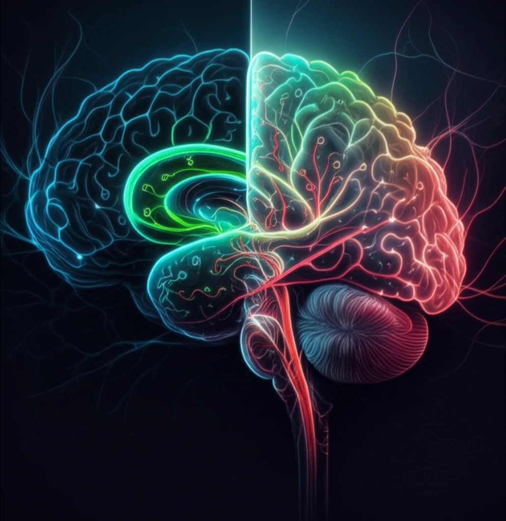
Decoding Fear: How Context Shapes Memory and Brain Structure
"New research uncovers dynamic changes in the brain's fear circuitry, offering insights into PTSD and anxiety disorders."
Fear is a fundamental emotion, essential for survival. It alerts us to danger, prompting a fight-or-flight response. However, when fear becomes detached from its original context, it can lead to debilitating conditions such as post-traumatic stress disorder (PTSD) and various anxiety disorders. Understanding how the brain processes and stores fear memories is crucial for developing effective treatments.
Contextual fear conditioning, a well-established model in neuroscience, provides a valuable framework for studying the formation of fear memories. This process involves associating a neutral context with an aversive stimulus, leading to a conditioned fear response when the context is encountered again. Researchers are delving deeper into the molecular mechanisms underlying this process to identify potential therapeutic targets.
A recent study published in the European Journal of Neuroscience sheds new light on the dynamic changes that occur in the brain during contextual fear conditioning. By employing advanced proteomic techniques, the researchers uncovered specific modifications in neuron and oligodendrocyte protein expression within the dentate gyrus, a critical region of the hippocampus involved in memory formation.
Unlocking the Brain's Fear Code: A Proteomic Analysis

The study, led by researchers at the Research Institute for Biosciences in Belgium, used an isotope-coded protein labeling (ICPL) approach to analyze protein expression changes in the dentate gyrus of rats 24 hours after contextual fear conditioning. This quantitative proteomic technique allowed the scientists to identify and measure a wide range of proteins involved in various cellular processes.
- Synaptic Plasticity: The study identified increased expression of proteins involved in vesicle trafficking, such as VAMP2 and RAB3C, suggesting enhanced synaptic communication in the dentate gyrus after fear conditioning.
- Neurite Outgrowth: Proteins that stimulate growth cone emergence and guidance, like BASP1 and calcineurin, were also upregulated, indicating active remodeling of neuronal connections.
- Myelin Structure: Intriguingly, the expression of myelin basic protein (MBP) and myelin proteolipid protein (PLP1) was decreased, suggesting a transient reduction in myelin in specific areas of the dentate gyrus.
Implications for Mental Health Treatment
This study provides valuable insights into the complex molecular mechanisms underlying fear memory formation. The discovery of dynamic changes in myelin structure, in particular, opens new avenues for research into the role of oligodendrocytes and myelin plasticity in learning and memory. By understanding how fear memories are encoded and consolidated in the brain, we can develop more targeted and effective treatments for PTSD and anxiety disorders, offering hope for those struggling with these debilitating conditions.
