Illustration of a horse's hind leg with ultrasound highlighting the tibial nerve.

Decoding Equine Lameness: A Guide to the Tibial Nerve Block

"Understanding and Utilizing Ultrasonography-Guided Nerve Blocks for Horses"


Lameness in horses can be a complex issue, often requiring a systematic approach to diagnose the root cause. Among the various diagnostic tools available, nerve blocks stand out as essential techniques for pinpointing the source of pain. The tibial nerve block, in particular, plays a crucial role in evaluating hindlimb lameness, allowing veterinarians to isolate the issue and develop targeted treatment plans.

Traditional blind injection methods, while commonly used, sometimes fall short in achieving optimal results. The anatomy of the tibial nerve and its surrounding structures can vary, leading to challenges in ensuring accurate and effective analgesia. This is where ultrasonography, or ultrasound imaging, steps in, offering a more precise and reliable solution.

This article delves into the application of ultrasonography-guided tibial nerve blocks. We'll explore the anatomical considerations, step-by-step techniques, and benefits of this advanced method. Whether you're a horse owner seeking to understand your equine companion's care or a veterinary professional looking to refine your skills, this guide provides valuable insights into this critical procedure.

Unveiling the Tibial Nerve: Anatomy and Ultrasonography

Illustration of a horse's hind leg with ultrasound highlighting the tibial nerve.

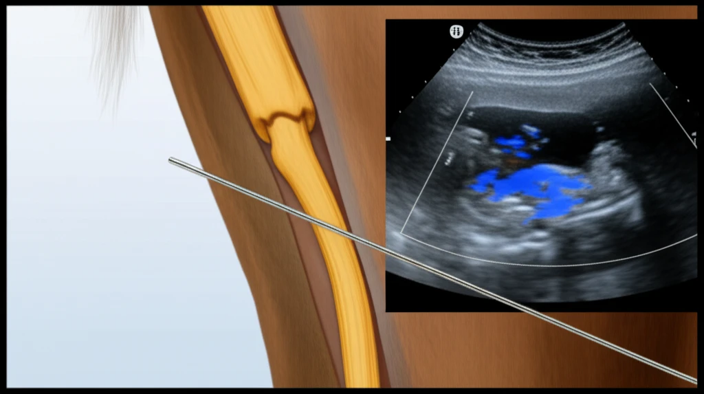
The tibial nerve, a major branch of the sciatic nerve, plays a pivotal role in hindlimb function. Understanding its anatomical pathway is fundamental to successful nerve blocking. The nerve runs along the caudal aspect of the femur and descends towards the hock, providing sensory and motor innervation to the lower limb.

Ultrasonography allows for real-time visualization of the tibial nerve and its surrounding structures. Using a microconvex probe, veterinarians can identify the nerve's location in the caudomedial aspect of the crus. The nerve appears as an oval structure, often nestled within a fatty layer, and can be visualized approximately 8-10 cm proximal to the point of the hock. This technique allows for precise needle placement and accurate anesthetic delivery.

  • Caudomedial Aspect: The tibial nerve is located in the inner, rear portion of the leg.
  • Oval Appearance: The nerve appears as an oval structure on an ultrasound.
  • Precise Location: Ultrasound helps locate the nerve 8-10 cm above the hock.
The process involves inserting a needle caudally to the probe, passing through the superficial caudal crural fascia. The needle is then guided towards the caudal aspect of the nerve, where the anesthetic solution is administered. A second injection is typically made cranial to the probe to ensure comprehensive coverage.

Enhancing Equine Care: The Benefits of Ultrasonography-Guided Nerve Blocks

Ultrasonography-guided tibial nerve blocks represent a significant advancement in equine veterinary medicine. By providing real-time visualization and precise needle placement, these techniques improve accuracy, reduce the risk of complications, and enhance the overall effectiveness of the nerve block. This approach not only benefits veterinary professionals but also contributes to improved outcomes and a higher quality of life for our equine companions.

About this Article -

This article was crafted using a human-AI hybrid and collaborative approach. AI assisted our team with initial drafting, research insights, identifying key questions, and image generation. Our human editors guided topic selection, defined the angle, structured the content, ensured factual accuracy and relevance, refined the tone, and conducted thorough editing to deliver helpful, high-quality information.See our About page for more information.

This article is based on research published under:

DOI-LINK: 10.1111/eve.13020, Alternate LINK

Title: Ultrasonographic Guided Block Of The Tibial Nerve

Subject: Equine

Journal: Equine Veterinary Education

Publisher: Wiley

Authors: J.‐M. Denoix, A. Beaumont, L. Bertoni

Published: 2018-10-30

Everything You Need To Know

1

What is a tibial nerve block, and why is it used in horses?

A tibial nerve block is a diagnostic technique used in equine veterinary medicine to identify the source of hindlimb lameness. It involves injecting a local anesthetic near the tibial nerve, a major branch of the sciatic nerve, to temporarily block pain signals from the lower limb. This procedure helps veterinarians pinpoint whether the lameness originates from structures innervated by the tibial nerve, aiding in the development of targeted treatment plans. The tibial nerve provides sensory and motor innervation to the lower limb.

2

How does ultrasonography improve the accuracy of a tibial nerve block?

Ultrasonography enhances the accuracy of a tibial nerve block by providing real-time visualization of the tibial nerve and its surrounding structures. This allows veterinarians to precisely identify the nerve's location in the caudomedial aspect of the crus, approximately 8-10 cm proximal to the hock. Using ultrasound, the needle placement is guided, ensuring the anesthetic solution is delivered directly to the nerve, thereby increasing the effectiveness of the block and reducing the risk of complications compared to traditional blind injection methods. The tibial nerve appears as an oval structure on the ultrasound.

3

Can you explain the step-by-step process of performing an ultrasonography-guided tibial nerve block?

The process begins with identifying the tibial nerve using a microconvex probe, which appears as an oval structure in the caudomedial aspect of the crus, 8-10 cm proximal to the hock. The veterinarian then inserts a needle caudally to the probe, passing through the superficial caudal crural fascia. The needle is guided towards the caudal aspect of the nerve, where the anesthetic solution is administered. Often, a second injection is made cranial to the probe to ensure comprehensive coverage of the nerve.

4

What are the key anatomical considerations when performing a tibial nerve block?

The primary anatomical consideration is the pathway of the tibial nerve, a major branch of the sciatic nerve, which runs along the caudal aspect of the femur and descends towards the hock. Understanding the nerve's location in the caudomedial aspect of the crus is crucial. Veterinarians must be aware of the nerve's position relative to the surrounding structures to ensure accurate needle placement and effective anesthesia. The use of ultrasonography aids in visualizing the nerve and its specific location.

5

What are the benefits of using ultrasonography-guided tibial nerve blocks for horses, compared to traditional methods?

Ultrasonography-guided tibial nerve blocks offer several advantages over traditional blind injection methods. They improve accuracy by allowing for precise needle placement, reducing the risk of complications because the veterinarian can visualize the anatomical structures. This results in enhanced effectiveness of the nerve block and can lead to better outcomes and a higher quality of life for the horse. It helps in developing more targeted treatment plans by accurately identifying the source of lameness.

Newsletter Subscribe

Subscribe to get the latest articles and insights directly in your inbox.