Decoding Embryo Development: A Multiscale Look at Axis Elongation
"Researchers quantify tissue behavior during amniote embryo axis elongation, revealing insights into morphogenesis."
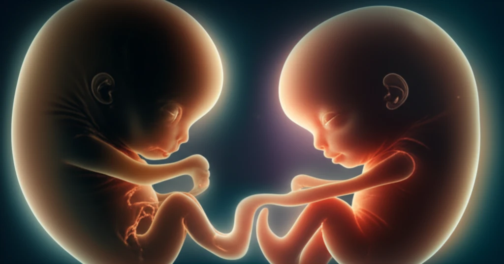
Embryonic development is a marvel of coordinated cellular activity, particularly the process of axis elongation, which lays the foundation for the body plan in amniotes. This intricate process involves multiple tissues working in harmony to form the posterior part of the developing organism. Understanding how these tissues communicate and coordinate their movements and growth has remained a significant challenge in developmental biology.
Traditional research methods have often fallen short in capturing the full complexity of these interactions. However, recent technological advancements in microscopy and image analysis are opening new avenues for studying morphogenesis at an unprecedented scale. These tools allow scientists to observe cellular behaviors in real-time and quantify the contributions of individual tissues to the overall developmental process.
A groundbreaking study has utilized these advanced techniques to investigate axis elongation in quail embryos. By combining high-resolution imaging, transgenic models, and sophisticated computational analysis, researchers have uncovered new insights into the tissue-specific behaviors and coordinated movements that drive this critical developmental event. This research not only enhances our understanding of fundamental biological processes but also holds promise for future applications in regenerative medicine and developmental therapies.
How Do Tissues Coordinate During Axis Elongation?
To unravel the complexities of axis elongation, researchers employed a multifaceted approach centered on quail embryos. Quail embryos are excellent models for studying amniote development due to their accessibility and the ability to visualize cellular processes in real-time. The team used transgenic quail embryos expressing fluorescent proteins, allowing them to track individual cells and tissues with remarkable precision.
- Confocal Microscopy and Live Imaging: Advanced microscopy techniques allowed for real-time observation of cellular movements and tissue interactions.
- Transgenic Quail Embryos: Fluorescent proteins enabled detailed tracking of cells and tissues.
- 3D Volumetric Techniques: Quantitative analysis of tissue volumes and cell densities provided insights into tissue-specific contributions.
- 4D Imaging and Image Analysis: Sophisticated algorithms were used to analyze cell motion and tissue deformations over time.
The Future of Understanding Embryonic Development
This research marks a significant step forward in our understanding of embryonic development, providing a quantitative and multiscale approach to analyzing tissue behaviors during morphogenesis. By integrating advanced imaging techniques with computational analysis, scientists can now dissect the complex interactions that drive axis elongation and other critical developmental processes. These insights pave the way for future investigations into the mechanisms of developmental disorders and the potential for regenerative therapies. Further studies could explore how genetic and environmental factors influence tissue coordination, potentially leading to new strategies for preventing birth defects and promoting tissue repair.