Decoding Dementia: How Brain Imaging is Revolutionizing Diagnosis and Treatment
"Unveiling the Future of Alzheimer's: Exploring the Cutting-Edge Role of Neuroimaging in Early Detection and Personalized Care."
Dementia, a term that evokes both fear and uncertainty, is a collection of symptoms characterized by a decline in cognitive abilities like memory, thinking, and judgment. While the condition affects millions worldwide, progress in diagnosis and treatment has been slow. But now, a revolution is underway, fueled by advancements in neuroimaging – the use of sophisticated brain scans to visualize the inner workings of the mind.
This groundbreaking technology is offering unprecedented insights into the structural and functional changes that occur in the brain during the progression of dementia, especially Alzheimer's disease (AD). Imagine being able to detect the earliest signs of the disease years before symptoms even appear. That's the promise of neuroimaging, and it's a promise that's rapidly becoming a reality.
This article dives deep into the world of neuroimaging, exploring how it's changing the landscape of dementia care. We'll examine how these advanced techniques are used to diagnose the disease, monitor its progression, and even guide the development of new therapies. We'll also explore the incredible potential of neuroimaging to unlock a deeper understanding of dementia and pave the way for a future where effective treatments are available to everyone.
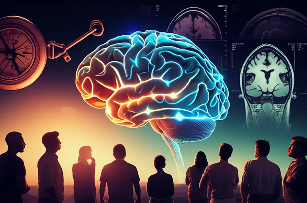
What is Neuroimaging and How Does it Work?
Neuroimaging encompasses a range of techniques that provide detailed pictures of the brain. These methods go far beyond the basic X-rays and CT scans that show the skull; they allow doctors and researchers to peer inside the brain to see its structure, function, and even its biochemical activity. Think of it like having a window into the brain, allowing you to observe what's happening in real-time.
- Magnetic Resonance Imaging (MRI): Uses powerful magnetic fields and radio waves to create detailed images of the brain's structure. MRI can reveal brain shrinkage (atrophy), which is a hallmark of many dementias.
- Positron Emission Tomography (PET): Uses radioactive tracers to visualize brain activity, such as glucose metabolism or the buildup of amyloid plaques, a key feature of Alzheimer's disease.
- Computed Tomography (CT): While less detailed than MRI, CT scans can be useful for ruling out other conditions that might mimic dementia symptoms, such as strokes or tumors.
- Single-Photon Emission Computed Tomography (SPECT): Similar to PET, SPECT uses radioactive tracers to assess brain blood flow, providing information about brain function.
The Future of Dementia Care: A Brighter Horizon
Neuroimaging is more than just a diagnostic tool; it's a catalyst for change. As technology advances and our understanding of the brain deepens, neuroimaging will play an increasingly vital role in the fight against dementia. From earlier, more accurate diagnoses to personalized treatment plans tailored to each individual, the future of dementia care looks brighter than ever before. By embracing these innovative tools, we are not just imaging the brain; we are imaging a future with hope.