Decoding DCM: What Canine Heart Changes Really Mean
"Unraveling Dilatative Cardiomyopathy: New Insights into Heart Health for Your Dog"
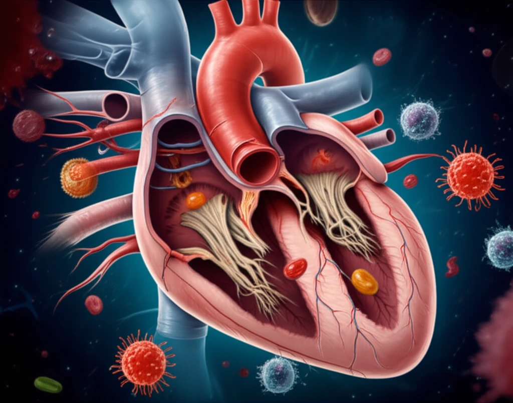
Dilatative cardiomyopathy (DCM) in dogs involves complex changes in the heart, including inflammation, fibrosis, and altered blood vessel development. While these changes often appear as localized problems, new research aims to understand the broader, diffuse changes occurring throughout the heart muscle of dogs with DCM.
A recent study used advanced imaging techniques to quantify key features in the hearts of dogs with DCM compared to healthy controls. This involved measuring cell counts, assessing the presence of specific immune cells (macrophages), and evaluating the amount of collagen and free space within the heart tissue.
By understanding these diffuse changes, researchers hope to gain insights into the underlying mechanisms driving DCM and potentially identify new targets for treatment. This article breaks down the study's findings, explaining what these heart changes mean for your dog and what future research may explore.
Key Changes in DCM-Affected Hearts
The study revealed several significant differences in the hearts of dogs with DCM compared to healthy dogs:
- Cardiomyocyte Reduction: The proportion of heart tissue occupied by cardiomyocytes was significantly less in dogs with DCM.
- Increased Collagen: There was a higher amount of collagen in the spaces between cells (interstitial space). Collagen is a fibrous protein that contributes to scarring, suggesting increased fibrosis in DCM hearts.
- Macrophage Infiltration: The number of macrophages, a type of immune cell, was elevated in the interstitial space. This indicates increased inflammation within the heart tissue.
- Interstitial Edema: The amount of free space in the interstitium was greater, suggesting fluid accumulation or edema.
- Vessel Density: The number of blood vessels in the heart tissue did not significantly change.
Implications and Future Directions
The study's findings suggest that DCM involves a combination of interstitial fibrosis, macrophage infiltration, and a potential increase in vascular permeability. These processes, along with the likely gradual loss of cardiomyocytes, appear to be key mechanisms driving the disease.
One critical question is whether these changes stem from an initial injury to the heart muscle cells themselves, followed by macrophage recruitment, or if macrophages are activated by a persistent inflammatory signal. Further research is needed to clarify this.
Ultimately, a deeper understanding of these mechanisms could pave the way for more targeted therapies to slow or prevent the progression of DCM in dogs, improving their quality of life and extending their lifespan.