Advanced imaging technology scanning a breast cancer cell.

Decoding Breast Cancer: How Advanced Imaging is Changing the Game

"Explore how PET scans, using cutting-edge radiopharmaceuticals, offer new insights into breast cancer, paving the way for personalized treatments."


Breast cancer's complexity arises from its heterogeneity, meaning each subtype exhibits unique molecular characteristics and progression patterns. This diversity underscores the need for advanced diagnostic tools that can accurately assess the specific features of an individual's cancer. Molecular imaging techniques, which provide functional information, play a vital role in evaluating treatment response and long-term prognosis.

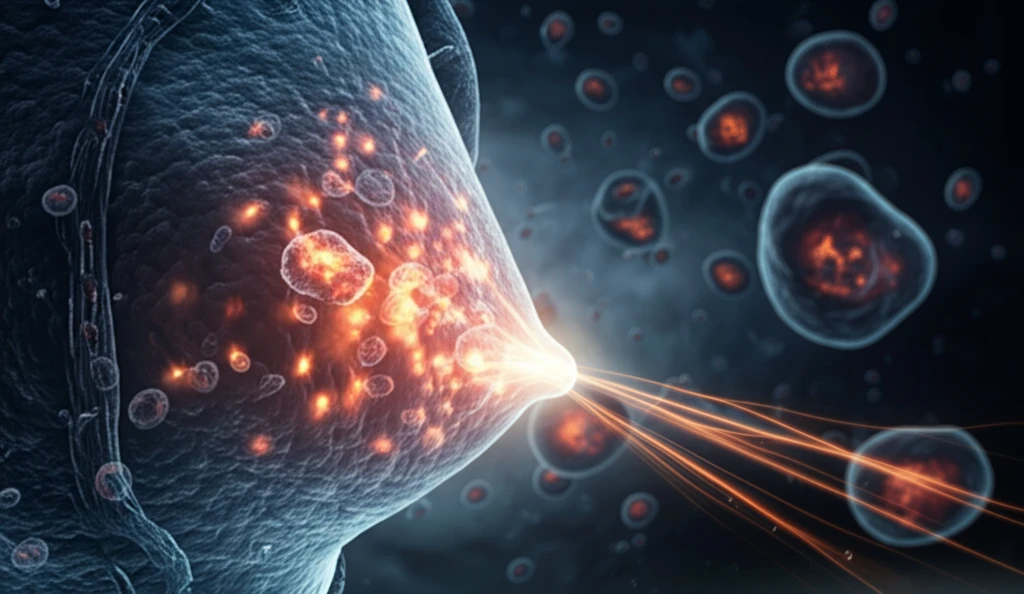
Nuclear imaging, including Positron Emission Tomography (PET), is essential for researching cancer biology and developing novel treatments. PET scans use radiopharmaceuticals to locate tumors, define their stage, and monitor their response to therapy.

This article explores the utility of 18F-fluorodeoxyglucose (18F-FDG) PET, the most widely used molecular imaging technique, and investigates the development of new radiopharmaceuticals like 18F-fluoro-17-estradiol (FES), 18F-fluoro-l-thymidine (FLT), 18F-fluoromisonidazole (FISO), and 89Zr-immuno-PET, which provide deeper insights into tumor characteristics.

How PET Scans are Revolutionizing Breast Cancer Management

Advanced imaging technology scanning a breast cancer cell.

PET scans offer a significant advantage over traditional imaging methods by visualizing biological processes at the cellular and molecular level. While guidelines advise against PET scans for initial staging in early breast cancer due to potential false negatives with small lesions, PET proves invaluable in complex cases.

PET imaging is recommended when conventional staging is unclear, particularly in advanced or metastatic disease. It excels at identifying unexpected regional or distant metastases and monitoring treatment response. Furthermore, PET scans can detect recurrence when tumor markers rise without visible signs on other imaging.

  • Accurate Staging: PET/CT scans enhance the accuracy of breast cancer staging, enabling healthcare providers to make well-informed treatment decisions.
  • Treatment Monitoring: Molecular imaging is crucial for assessing how breast cancer responds to therapies such as chemotherapy. This helps doctors adapt strategies quickly.
  • Personalized Medicine: PET scans can help individualize breast cancer treatment by revealing particular molecular characteristics, which could improve results.
Dual-point FDG imaging enhances accuracy by differentiating malignant from benign lesions through changes in FDG uptake over time. This technique involves imaging lesions at multiple time points to monitor changes in SUV, helping distinguish between cancerous and non-cancerous conditions.

The Future of Breast Cancer Imaging

Combining PET imaging with techniques that capture critical biological events—ER, PR, and HER2 expression, angiogenesis, and metabolic changes—holds the key to improving diagnosis and treatment. Ongoing studies aim to refine the specificity and utility of radiopharmaceuticals, paving the way for truly personalized medicine where treatment selection is guided by the unique characteristics of each patient's tumor.

About this Article -

This article was crafted using a human-AI hybrid and collaborative approach. AI assisted our team with initial drafting, research insights, identifying key questions, and image generation. Our human editors guided topic selection, defined the angle, structured the content, ensured factual accuracy and relevance, refined the tone, and conducted thorough editing to deliver helpful, high-quality information.See our About page for more information.

This article is based on research published under:

DOI-LINK: 10.1186/s41824-018-0039-x, Alternate LINK

Title: Positron Emission Tomography In Breast Cancer: 18F- Fdg And Other Radiopharmaceuticals

Subject: Radiology, Nuclear Medicine and imaging

Journal: European Journal of Hybrid Imaging

Publisher: Springer Science and Business Media LLC

Authors: Ioannis Iakovou, Evanthia Giannoula, Areti Gkantaifi, Sofia Levva, Savvas Frangos

Published: 2018-10-24

Everything You Need To Know

1

What is the role of PET scans in diagnosing and treating breast cancer?

PET scans play a crucial role in breast cancer management by visualizing biological processes at the cellular and molecular level. They are used for accurate staging, particularly in advanced or metastatic disease, where conventional staging might be unclear. PET scans help identify unexpected metastases and monitor treatment response. In addition, they can detect cancer recurrence when tumor markers rise, even if other imaging methods show no visible signs. PET's ability to assess the specific features of an individual's cancer contributes to personalized treatment strategies. The use of radiopharmaceuticals in PET scans, like 18F-FDG and others such as 18F-FES, 18F-FLT, 18F-FISO, and 89Zr-immuno-PET, provides detailed insights into tumor characteristics, which helps guide therapy decisions.

2

How do PET scans compare to traditional imaging methods in breast cancer detection?

PET scans offer a significant advantage over traditional imaging methods because they can visualize biological processes at the cellular and molecular level. Unlike traditional methods, PET scans utilize radiopharmaceuticals to provide functional information, which is crucial for evaluating treatment response and long-term prognosis. While traditional imaging methods may be sufficient for initial staging in early breast cancer, PET imaging is more effective in complex cases, such as advanced or metastatic disease, where it excels at identifying unexpected regional or distant metastases and monitoring treatment response. PET scans are also recommended when conventional staging is unclear.

3

Can you explain the significance of different radiopharmaceuticals mentioned in the context of breast cancer imaging?

Several radiopharmaceuticals are discussed in this context, each providing unique insights into breast cancer. 18F-fluorodeoxyglucose (18F-FDG) is the most widely used molecular imaging technique in PET scans, helping to locate tumors and monitor their response to therapy. Other radiopharmaceuticals, such as 18F-fluoro-17-estradiol (FES), 18F-fluoro-l-thymidine (FLT), 18F-fluoromisonidazole (FISO), and 89Zr-immuno-PET, offer deeper insights into specific tumor characteristics. For instance, 18F-FES targets estrogen receptors, aiding in assessing hormone receptor status, which is vital for treatment planning. Understanding the roles of these radiopharmaceuticals is essential for personalized cancer care, as they enable the tailoring of treatment strategies to match the unique characteristics of each patient's tumor.

4

How does dual-point FDG imaging enhance the accuracy of PET scans?

Dual-point FDG imaging enhances the accuracy of PET scans by differentiating between malignant and benign lesions through changes in FDG uptake over time. This technique involves imaging lesions at multiple time points to monitor changes in the Standardized Uptake Value (SUV). By observing how FDG uptake changes, healthcare providers can better distinguish between cancerous and non-cancerous conditions. This method improves the specificity of PET scans, helping to avoid false positives and negatives, thereby leading to more accurate diagnoses and treatment decisions. This approach is a valuable tool for improving the reliability of PET scans in breast cancer detection and management.

5

What is the future of breast cancer imaging and how can it improve personalized medicine?

The future of breast cancer imaging involves combining PET imaging with techniques that capture critical biological events, such as ER, PR, and HER2 expression, angiogenesis, and metabolic changes. This integrated approach aims to enhance both the diagnosis and treatment of breast cancer. Ongoing studies are focused on refining the specificity and utility of radiopharmaceuticals. By understanding the unique characteristics of each patient's tumor at a molecular level, doctors can tailor treatment strategies to optimize outcomes. The goal is to move towards truly personalized medicine, where treatment selection is guided by the specific features of each patient's cancer, leading to more effective therapies and improved patient outcomes. The development of new radiopharmaceuticals, like 18F-FES, 18F-FLT, 18F-FISO, and 89Zr-immuno-PET, plays a key role in this evolution.

Newsletter Subscribe

Subscribe to get the latest articles and insights directly in your inbox.