Breast ultrasound and microscopic tissue aligning, symbolizing accurate diagnosis.

Decoding Breast Biopsy Results: How to Ensure Accuracy and Peace of Mind

"Understanding imaging-pathology concordance for accurate breast health diagnosis and management after ultrasound-guided biopsy."


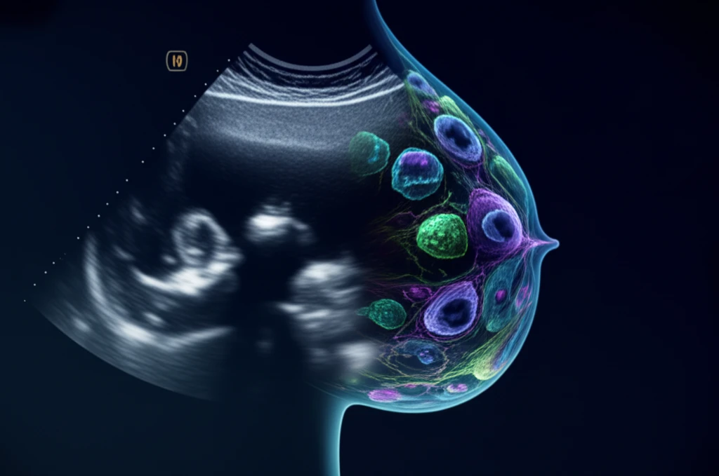
Ultrasound (US)-guided breast biopsy has become a cornerstone in diagnosing breast conditions due to its high diagnostic accuracy, approaching the reliability of open surgical biopsies. However, confirming adequate tissue retrieval after a US-guided biopsy remains a challenge, making false-negative results a possibility.

A critical step in validating biopsy results and guiding appropriate patient care is determining imaging-pathology concordance – essentially, ensuring that what the images show aligns with what the pathology report reveals. This process helps to minimize errors and ensures patients receive the correct diagnosis and treatment plan.

This article will explore the results of US-guided breast biopsies, discuss key considerations for establishing imaging-pathology concordance, review various categories of imaging-pathology correlations, and outline appropriate management strategies for each scenario. Understanding these elements can empower patients and healthcare providers to make informed decisions.

What is Imaging-Pathology Concordance and Why Does it Matter?

Breast ultrasound and microscopic tissue aligning, symbolizing accurate diagnosis.

Imaging-pathology concordance refers to the agreement between the findings on breast imaging (such as ultrasound) and the results of the pathology analysis performed on the tissue sample obtained during a biopsy. When the imaging and pathology results align, it strengthens the confidence in the diagnosis.

However, discrepancies can occur, leading to what is known as imaging-pathology discordance. This can happen for several reasons:

  • Inadequate Targeting: The biopsy needle may not have accurately targeted the suspicious area seen on the imaging.
  • Sampling Error: The tissue sample obtained may not be representative of the entire lesion, especially if the lesion is heterogeneous.
  • Technical Difficulties: Poor visualization of the lesion or needle during the procedure, dense breast tissue, or lesion mobility can hinder accurate targeting.
  • Interpretative Errors: Discrepancies can arise from errors in interpreting either the imaging findings or the pathology results.
Accurate interpretation of both imaging and pathology is crucial. Radiologists must be familiar with the Breast Imaging Reporting and Data System (BI-RADS) lexicon and classification to accurately assess lesions and guide biopsy procedures. When discrepancies arise, careful review and communication between the radiologist and pathologist are essential to determine the best course of action.

Ensuring Accuracy: A Collaborative Approach

Given the potential for discordance and its impact on patient care, a collaborative approach is crucial. This involves open communication between the radiologist performing the biopsy, the pathologist interpreting the tissue sample, the referring physician, and the patient.

When imaging-pathology discordance is suspected, the team should discuss the case, review the images and pathology slides, and consider whether additional tissue sampling is needed. This may involve a repeat biopsy using a different technique, such as vacuum-assisted breast biopsy (VABB), or surgical excision for a more comprehensive evaluation.

Ultimately, the goal is to ensure an accurate diagnosis and appropriate management plan, providing patients with the best possible care and peace of mind.

About this Article -

This article was crafted using a human-AI hybrid and collaborative approach. AI assisted our team with initial drafting, research insights, identifying key questions, and image generation. Our human editors guided topic selection, defined the angle, structured the content, ensured factual accuracy and relevance, refined the tone, and conducted thorough editing to deliver helpful, high-quality information.See our About page for more information.

This article is based on research published under:

DOI-LINK: 10.14366/usg.17049, Alternate LINK

Title: Evaluating Imaging-Pathology Concordance And Discordance After Ultrasound-Guided Breast Biopsy

Subject: Radiology, Nuclear Medicine and imaging

Journal: Ultrasonography

Publisher: Korean Society of Ultrasound in Medicine

Authors: Vivian Youngjean Park, Eun-Kyung Kim, Hee Jung Moon, Jung Hyun Yoon, Min Jung Kim

Published: 2018-04-01

Everything You Need To Know

1

What exactly is imaging-pathology concordance, and why is it so crucial in breast health diagnosis?

Imaging-pathology concordance is the agreement between what's seen on breast imaging, like ultrasound, and what the pathology analysis reveals from the tissue sample taken during a biopsy. It's important because when the imaging and pathology results align, it increases confidence in the diagnosis, leading to more appropriate treatment plans.

2

What are some reasons why imaging-pathology discordance might occur after a breast biopsy, and how can it be addressed?

Discrepancies between imaging and pathology results, known as imaging-pathology discordance, can arise due to inadequate targeting during the biopsy, sampling errors where the tissue sample isn't representative of the entire lesion, technical difficulties during the procedure, or interpretative errors in either the imaging findings or the pathology results. Addressing discordance requires a collaborative approach involving radiologists and pathologists to review findings and determine the best course of action.

3

Why are false-negative results a possibility after an ultrasound-guided biopsy, and how does ensuring imaging-pathology concordance help?

False-negative results after a US-guided biopsy are possible because confirming adequate tissue retrieval can be challenging. This means that despite the biopsy, the results might incorrectly suggest the absence of a condition, such as cancer, when it is actually present. Ensuring imaging-pathology concordance helps minimize these errors by verifying that the tissue sample analyzed corresponds to the area of concern identified in the imaging.

4

What is the Breast Imaging Reporting and Data System (BI-RADS), and why is it so important for radiologists to be familiar with it?

The Breast Imaging Reporting and Data System (BI-RADS) lexicon and classification is a standardized system used by radiologists to assess breast lesions and guide biopsy procedures. Familiarity with BI-RADS is crucial for accurate interpretation of imaging findings. It ensures consistency in reporting and helps in making informed decisions about patient management. A radiologist uses BI-RADS to communicate findings with other physicians.

5

Why is a collaborative approach so important in ensuring the accuracy of breast biopsy results and subsequent treatment decisions?

A collaborative approach involving open communication between the radiologist, pathologist, referring physician, and the patient is crucial to minimize errors and ensure accurate diagnosis and treatment. This collaboration facilitates thorough review of imaging and pathology results, discussion of any discrepancies, and development of an appropriate management strategy. When discrepancies are found, the radiologist and the pathologist work together to get to the right diagnosis.

Newsletter Subscribe

Subscribe to get the latest articles and insights directly in your inbox.