AI brain tumor prediction model based on radiomics.

Decoding Brain Tumors: How AI Radiomics Can Predict Survival

"A non-invasive approach to predicting progression-free survival in lower-grade gliomas using radiomic signatures and genetic insights."


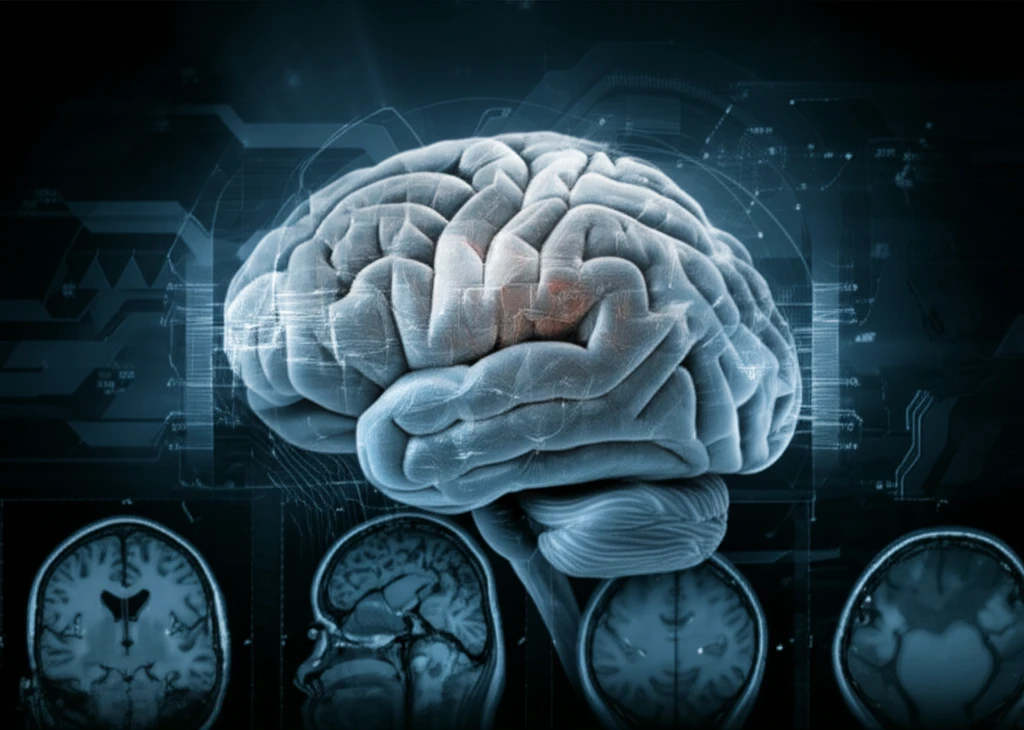
Gliomas, the most common primary tumors in the central nervous system, present a significant challenge due to their variable behavior and unpredictable progression. Lower-grade gliomas (LGGs), which include grade II and III gliomas, account for a substantial portion of these cases. Predicting how these tumors will progress is critical for effective treatment planning, yet current methods often fall short.

Traditional methods rely on clinical assessments and pathological analysis, but these can be invasive and may not fully capture the complex nature of LGGs. The emerging field of radiomics offers a promising alternative. Radiomics involves extracting a large number of quantitative features from medical images, such as MRI scans, and using these features to build predictive models.

This article explores groundbreaking research that uses AI to analyze MRI scans of LGGs, creating a 'radiomic signature' capable of predicting progression-free survival (PFS). By combining radiomics with genetic analysis, this approach provides a non-invasive and individualized assessment of tumor behavior, potentially transforming how LGGs are diagnosed and treated.

AI-Powered Insights: Predicting LGG Progression with Radiomics

AI brain tumor prediction model based on radiomics.

Researchers from Capital Medical University in Beijing have developed a novel method for predicting the progression of lower-grade gliomas (LGGs) using radiomics. This approach involves using artificial intelligence (AI) to analyze MRI scans and identify patterns that correlate with how the tumor is likely to behave. Here’s a breakdown of how the study was conducted:

The study involved analyzing MRI scans from 216 patients to train the AI, then validating it on a separate group of 84 patients. The AI extracted 431 different quantitative features from the MRI images. These features included:

  • First-order statistics: Describing the distribution of signal intensity in the images.
  • Shape- and size-based features: Quantifying the tumor's form.
  • Textural features: Reflecting the heterogeneity within the tumor.
  • Wavelet features: Derived from the above features using wavelet decomposition.
The AI then identified a specific combination of nine radiomic features that were most strongly associated with progression-free survival (PFS). This combination is the 'radiomic signature.' The study found that patients could be classified into high-risk and low-risk groups. Using this radiomic signature, the accuracy of prognosis was significantly improved and validated by genetic analysis.

The Future of Brain Tumor Treatment: Personalized and Non-Invasive

This research marks a significant step forward in the treatment of LGGs. By using AI to analyze MRI scans, doctors can gain a more accurate understanding of how a tumor is likely to progress, enabling more personalized treatment plans.

The radiogenomic analysis further enhances this approach by linking the radiomic signature to specific biological processes, such as immune response and cell proliferation. This opens the door for targeted therapies that address the unique characteristics of each tumor.

While further studies are needed to validate these findings, the potential impact of radiomics on brain tumor treatment is undeniable. This non-invasive approach promises to improve patient outcomes and transform the way LGGs are managed in the future.

About this Article -

This article was crafted using a human-AI hybrid and collaborative approach. AI assisted our team with initial drafting, research insights, identifying key questions, and image generation. Our human editors guided topic selection, defined the angle, structured the content, ensured factual accuracy and relevance, refined the tone, and conducted thorough editing to deliver helpful, high-quality information.See our About page for more information.

This article is based on research published under:

DOI-LINK: 10.1016/j.nicl.2018.10.014, Alternate LINK

Title: A Radiomic Signature As A Non-Invasive Predictor Of Progression-Free Survival In Patients With Lower-Grade Gliomas

Subject: Cognitive Neuroscience

Journal: NeuroImage: Clinical

Publisher: Elsevier BV

Authors: Xing Liu, Yiming Li, Zenghui Qian, Zhiyan Sun, Kaibin Xu, Kai Wang, Shuai Liu, Xing Fan, Shaowu Li, Zhong Zhang, Tao Jiang, Yinyan Wang

Published: 2018-01-01

Everything You Need To Know

1

What exactly does radiomics involve in the context of brain tumor analysis?

Radiomics involves extracting a large number of quantitative features from medical images like MRI scans. These features are then used to build predictive models, offering a non-invasive method to understand tumor characteristics. This approach contrasts with traditional methods that often rely on invasive procedures like biopsies for pathological analysis. Radiomics captures the complex nature of lower-grade gliomas (LGGs) by quantifying various aspects visible in the images.

2

How does artificial intelligence (AI) analyze MRI scans to predict the progression of lower-grade gliomas (LGGs)?

The AI analyzes MRI scans to identify a 'radiomic signature' that correlates with progression-free survival (PFS) in lower-grade gliomas (LGGs). It extracts hundreds of quantitative features, including first-order statistics (signal intensity), shape- and size-based features (tumor form), textural features (heterogeneity), and wavelet features. By combining a specific set of these features, the AI can classify patients into high-risk and low-risk groups.

3

What is the significance of identifying a 'radiomic signature' in predicting the behavior of lower-grade gliomas (LGGs)?

The study identified a specific combination of nine radiomic features that were strongly associated with progression-free survival (PFS) in lower-grade gliomas (LGGs). This combination, known as the 'radiomic signature', enables the classification of patients into different risk groups. This risk stratification helps doctors understand how a tumor is likely to behave and progress, which is crucial for effective treatment planning.

4

How does genetic analysis enhance the predictive power of radiomics in assessing lower-grade gliomas (LGGs)?

The integration of genetic analysis with radiomics enhances the accuracy of prognosis for lower-grade gliomas (LGGs). By validating the radiomic signature with genetic data, researchers can confirm the reliability of the AI-driven predictions. This combined approach provides a more comprehensive and individualized assessment of tumor behavior, improving the confidence in treatment decisions.

5

What are the implications of AI-driven radiomics for personalized treatment of lower-grade gliomas (LGGs), and why is this significant?

This research represents a significant advancement in personalized medicine for lower-grade gliomas (LGGs). By using AI and radiomics, treatment plans can be tailored to the individual patient's tumor characteristics and predicted progression. This approach can lead to more effective treatments, improved survival rates, and a better quality of life for patients. Personalized approaches contrast with traditional methods that may not fully capture the unique aspects of each patient's condition.

Newsletter Subscribe

Subscribe to get the latest articles and insights directly in your inbox.