Decoding Brain Changes After Heart Surgery: What Every Patient Should Know
"A new study sheds light on how CT scans can help identify and manage neurological complications following cardiovascular procedures."
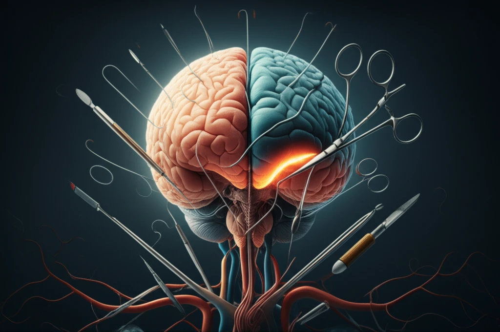
Cardiovascular surgeries, while life-saving, carry a risk of neurological complications (NC). These complications can significantly impact a patient's recovery and long-term well-being, leading to increased morbidity and mortality. Understanding the causes and being able to quickly identify these issues is critical for effective management and improved patient outcomes.
A recent study investigated the use of non-contrast computed tomography (NCCT) scans in detecting neurological problems that arise shortly after cardiovascular surgery. The aim was to not only confirm the utility of NCCT, but also to identify the spectrum of brain changes associated with different types of cardiovascular procedures.
This article breaks down the key findings of the study, explaining what these brain changes look like on CT scans, how they relate to different surgeries, and what it all means for patients and their care.
What Brain Changes Can Happen After Heart Surgery?
The study, which retrospectively analyzed NCCT scans of patients who experienced neurological issues within seven days of cardiovascular surgery, revealed that a significant portion – 6.7% – developed neurological complications. These complications ranged from obvious stroke-like symptoms (focal deficits) to more subtle changes like seizures, delirium, or cognitive impairment (non-focal deficits).
- Stroke vs. Non-Focal Deficits: Patients with stroke-like symptoms were more likely to have positive findings on their CT scans compared to those with non-focal deficits.
- Positive CT Scan Findings: A large majority (88%) of patients with neurological complications had positive findings on their CT scans. The most common findings were ischemic infarcts (areas of brain tissue damage due to lack of blood flow), followed by subdural hemorrhages (bleeding between the brain and its outer covering). Other findings included intra-parenchymal hemorrhages, sub-arachnoid hemorrhages, and cerebro-vascular thrombosis.
- Pediatric Considerations: Neurological complications also occurred in children undergoing surgery for complex congenital heart defects.
The Future of Post-Operative Neurological Care
This study reinforces the value of NCCT scans as a readily available and effective tool for diagnosing neurological complications after cardiovascular surgery. The ability to quickly visualize brain changes allows medical teams to make informed decisions about treatment and management.
While NCCT is valuable, it's important to remember that it's just one piece of the puzzle. Clinical evaluation and other diagnostic tests are also crucial for a comprehensive assessment. For instance, in a small percentage of patients in the study (11%), NCCT scans were normal despite neurological symptoms.
Ultimately, this research contributes to the ongoing effort to improve patient outcomes after cardiovascular surgery by highlighting the importance of early neurological monitoring and the role of imaging technologies in guiding treatment strategies. As medical technology evolves, we can expect even more precise and personalized approaches to managing post-operative neurological health.