A detailed digital illustration of a human radius bone cross-section, showcasing cortical porosity and a healthy bone structure.

Decoding Bone Health: How Modern Science Measures Your Risk and What You Can Do About It

"New research unveils advanced techniques for assessing bone porosity, offering insights into osteoporosis and fracture risk."


Our bones, the silent guardians of our physical well-being, often go unnoticed until a fracture or a diagnosis of osteoporosis casts a spotlight on their importance. Bone health is not merely about the structural integrity of our skeletons; it's a dynamic process that reflects our overall health and lifestyle. In recent years, advancements in medical imaging have provided unprecedented opportunities to delve deeper into the intricacies of bone structure, offering new ways to assess risk and guide interventions.

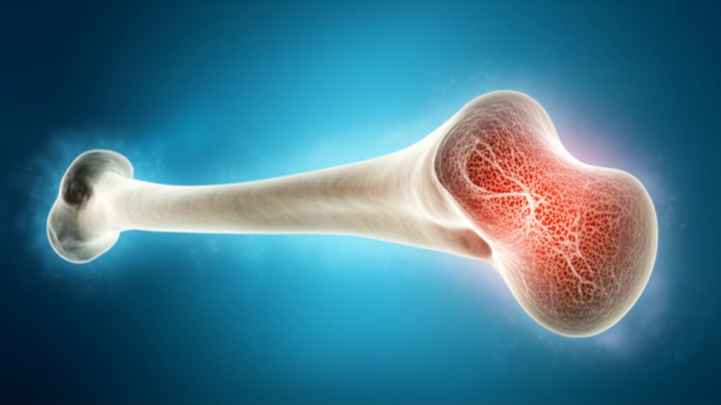
This article explores the latest research in bone health assessment, focusing on a study that compares different methods for measuring cortical porosity. Cortical porosity, or the presence of holes in the outer layer of bone, is a critical indicator of bone strength and fracture risk. By understanding how scientists measure porosity and interpret the results, you can gain valuable insights into your own bone health and make informed decisions about your care.

The study, published in Bone, examines the agreement between high-resolution peripheral quantitative computed tomography (HR-pQCT) and synchrotron radiation micro-CT (SR-µCT). HR-pQCT is a non-invasive imaging technique, while SR-µCT is a more advanced, laboratory-based method. By comparing these techniques, researchers are gaining a better understanding of how to accurately assess bone health and predict fracture risk.

Unveiling the Techniques: HR-pQCT and SR-µCT in Detail

A detailed digital illustration of a human radius bone cross-section, showcasing cortical porosity and a healthy bone structure.

The research hinges on two primary imaging techniques: HR-pQCT and SR-µCT. HR-pQCT, which is often used in clinical settings, offers a convenient, non-invasive way to assess bone structure. It works by using X-rays to create detailed images of the bone's architecture, including the cortical porosity in the distal radius, the lower part of your forearm.

In contrast, SR-µCT is a more sophisticated laboratory-based technique. It uses synchrotron radiation, a powerful form of X-ray, to generate high-resolution images. This allows researchers to see the bone's structure in incredible detail, essentially acting as a 'gold standard' for assessing bone health. However, SR-µCT is not available for routine clinical use.

  • HR-pQCT: A non-invasive technique using X-rays to create detailed images of the bone's structure.
  • SR-µCT: A laboratory-based technique using synchrotron radiation to generate high-resolution images.
  • Cortical Porosity: The presence of holes in the outer layer of bone, a key indicator of bone strength and fracture risk.
The study focused on the distal radius, a common site for fractures, particularly in older adults. By comparing the measurements obtained from HR-pQCT and SR-µCT, researchers can evaluate the accuracy of HR-pQCT in assessing bone health. The goal is to improve the way bone health is assessed in order to predict and prevent fractures.

Empowering Your Bone Health: Moving Forward

As research in bone health continues to evolve, so does our ability to safeguard our skeletal system. Understanding the different methods for assessing bone health, as well as the factors that influence it, is the first step in taking proactive steps towards a stronger, healthier future. Whether you're already concerned about your bone health or simply looking to maintain it, the insights from studies like these can help you make informed decisions. Consult with your healthcare provider to discuss your individual risk factors and explore strategies to keep your bones strong and resilient, for years to come.

About this Article -

This article was crafted using a human-AI hybrid and collaborative approach. AI assisted our team with initial drafting, research insights, identifying key questions, and image generation. Our human editors guided topic selection, defined the angle, structured the content, ensured factual accuracy and relevance, refined the tone, and conducted thorough editing to deliver helpful, high-quality information.See our About page for more information.

This article is based on research published under:

DOI-LINK: 10.1016/j.bone.2018.12.008, Alternate LINK

Title: Cortical Porosity Assessment In The Distal Radius: A Comparison Of Hr-Pqct Measures With Synchrotron-Radiation Micro-Ct-Based Measures

Subject: Histology

Journal: Bone

Publisher: Elsevier BV

Authors: Nikoo Soltan, Chantal E. Kawalilak, David M. Cooper, Saija A. Kontulainen, James D. Johnston

Published: 2019-03-01

Everything You Need To Know

1

What is cortical porosity, and why is it important for bone health?

Cortical porosity refers to the presence of holes in the outer layer of bone. It's a critical indicator of bone strength because increased porosity weakens the bone structure, making it more susceptible to fractures. Measuring cortical porosity helps in assessing overall bone health and predicting fracture risk, particularly in areas like the distal radius.

2

What are HR-pQCT and SR-µCT, and how do they differ in assessing bone structure?

HR-pQCT (high-resolution peripheral quantitative computed tomography) and SR-µCT (synchrotron radiation micro-CT) are both imaging techniques used to assess bone structure. HR-pQCT is a non-invasive method commonly used in clinical settings that uses X-rays to create detailed images of bone architecture, including cortical porosity. SR-µCT, on the other hand, is a laboratory-based technique that uses synchrotron radiation to generate very high-resolution images, providing a more detailed view of bone structure. While SR-µCT is considered a 'gold standard' due to its precision, it's not available for routine clinical use like HR-pQCT.

3

Why is the distal radius specifically studied in bone health research?

The distal radius, the lower part of your forearm, is a common site for fractures, especially in older adults. Because of this higher fracture incidence, it serves as a relevant area for studying bone health and the effectiveness of different measurement techniques like HR-pQCT and SR-µCT. Research on the distal radius helps in understanding how bone structure and porosity correlate with fracture risk in a clinically significant area.

4

How can understanding the measurements of cortical bone porosity using HR-pQCT and SR-µCT empower individuals to take proactive steps regarding their bone health?

Understanding how cortical porosity is measured using techniques like HR-pQCT and SR-µCT allows individuals to be more informed about their bone health status. By knowing the capabilities and limitations of each method, individuals can better interpret the results of bone density scans and have more meaningful discussions with their healthcare providers about their fracture risk. This knowledge empowers them to make informed decisions regarding lifestyle modifications, dietary changes, or medical interventions to strengthen their bones and reduce their risk of fractures.

5

What are the implications of research comparing HR-pQCT and SR-µCT for improving bone health assessment and fracture prevention?

Research that compares HR-pQCT and SR-µCT helps validate the accuracy and reliability of HR-pQCT, which is more accessible for clinical use. If HR-pQCT measurements are shown to correlate well with the 'gold standard' SR-µCT, clinicians can have greater confidence in using HR-pQCT to assess bone health and predict fracture risk. This can lead to earlier identification of individuals at risk of fractures, allowing for timely interventions such as lifestyle changes or medication to improve bone strength and prevent fractures. Further, understanding the nuances between these techniques can guide the development of even more accurate and accessible bone assessment tools in the future.

Newsletter Subscribe

Subscribe to get the latest articles and insights directly in your inbox.