
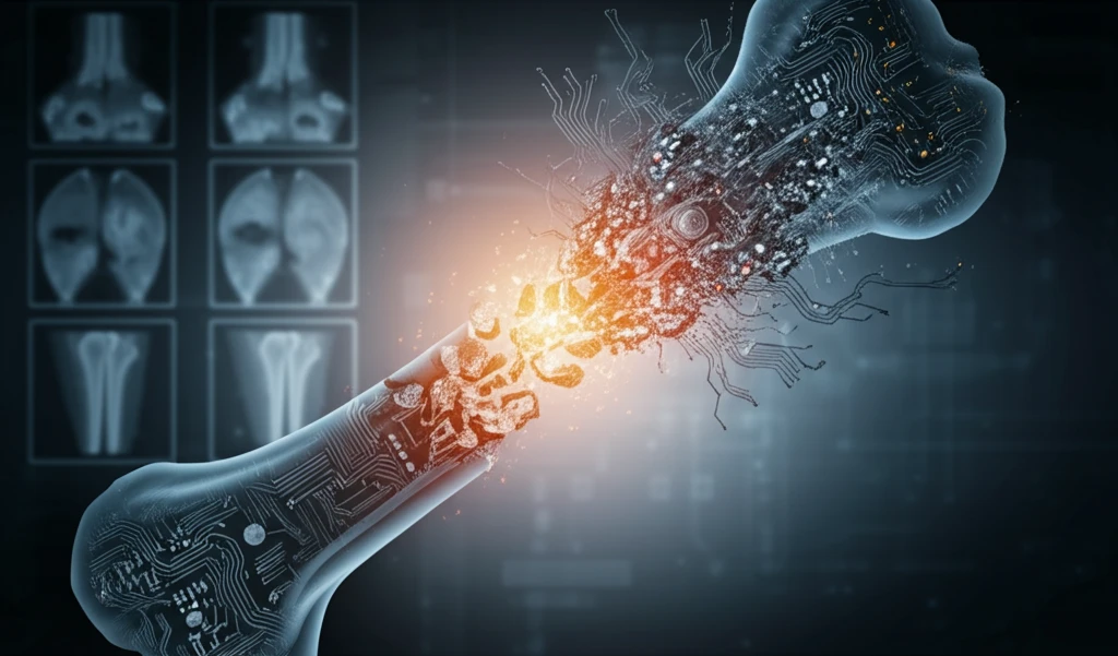
Decoding Bone Health: Can AI-Powered Image Analysis Revolutionize Osteoporosis Detection?

"Explore how cutting-edge image analysis techniques, fueled by artificial intelligence, are enhancing the precision and speed of osteoporosis diagnosis."
Osteoporosis, a condition characterized by decreased bone mass and increased fracture risk, affects millions worldwide, particularly postmenopausal women. Early and accurate diagnosis is crucial for effective management and prevention of severe consequences such as hip fractures. Traditional diagnostic methods, such as bone densitometry, have limitations, highlighting the need for innovative approaches.
The convergence of artificial intelligence (AI) and medical imaging is opening new frontiers in disease detection and management. AI-powered image analysis can enhance the precision and efficiency of diagnosing various conditions, including osteoporosis. By leveraging machine learning algorithms, subtle patterns and indicators within medical images can be detected, often missed by the human eye.
This article delves into how fractional Brownian motion (fBm), a mathematical concept used to describe natural phenomena, is being harnessed alongside AI to improve osteoporosis diagnosis. We'll explore how AI algorithms classify images generated by fBm, the effectiveness of these methods, and their potential to transform bone health assessments.
The Burden of Osteoporosis and the Case for Early Detection
Bone deterioration from osteoporosis and its resulting fractures primarily affect post-menopausal women and older adults, making the disease a significant public health concern. Early detection is considered very important because current diagnostic systems are both expensive and not universally accessible, which drives the search for more affordable screening methods. Researchers have explored using X-ray images of the vertebra—for example, through blended statistical methods and the Index-Singh classification—to identify bone density loss. Screening guidance is population-specific; the U.S. Preventive Services Task Force, for instance, asks clinicians to weigh the potential preventable burden and cites age-adjusted National Health and Nutrition Examination Survey data from 2017 to 2018 when deciding whether to screen men.
Gold-Standard DXA and the Limits of Current Detection Methods
Dual-energy X-ray absorptiometry (DXA/DEXA) remains the clinical gold standard for measuring bone mineral density, a consensus reflected in imaging reviews and clinical summaries alike. Conventional detection relies primarily on imaging and laboratory approaches to determine bone mineral density and bone quality, with computed tomography among the supporting modalities—though each imaging technique carries distinct advantages and limitations. These established methods are not without shortcomings: they can be expensive and less accessible, and AI classification research in this space frequently contends with small or imbalanced datasets that restrict model generalizability and increase the risk of overfitting. As a result, clinicians still face real barriers to timely, affordable diagnosis.
From Silent Disease to Detectable Condition: Foundations of Osteoporosis Screening
Osteoporosis is widely described as a silent disease: typically there are no symptoms unless a person breaks a bone, at which point the fracture can cause significant pain. The bone mineral density test—which examines segments of bone through X-rays and measures calcium and other minerals in the bones—became the cornerstone of diagnosis, with T-scores indicating the severity of bone loss. Long-standing clinical understanding holds that age, gender, family history, and ethnicity all shape disease risk, which is why family-history questions, such as whether a parent has had a hip fracture, remain central to assessment. Recognized early warning signs include low bone density, bone fractures, lost height, a curved upper back, sudden back pain, gastrointestinal issues, and dental problems.
AI and Fractional Brownian Motion: A New Diagnostic Era
Fractional Brownian motion (fBm) is a mathematical concept used to characterize various natural phenomena, from landscapes to stock market fluctuations. In medical imaging, fBm helps model and analyze complex textures, such as those found in bone structures. By synthesizing images using fBm, researchers can create models that mimic the intricate patterns of bone tissue, allowing for better assessment of bone health.
- Dual-tree MBand Decomposition Transform (DMBDT): Extracts crucial texture features from the synthesized images, providing a detailed multi-scale analysis.
- Support Vector Machine (SVM) Classifier: Classifies the images based on the extracted features, evaluating the quality and accuracy of the fBm synthesis methods.
- Statistical Feature Analysis: Includes measures such as mean, variance, mode, and Renyi entropy to quantify the characteristics of bone texture.
Machine Learning, Deep Learning, and Radiomics Enter the Clinic
Machine learning is changing how osteoporosis is detected, with a recent integrative review taking a step-by-step view of the technology and providing insights into today's detection landscape and the outlook for the future. A systematic review and meta-analysis in the Journal of Medical Internet Research evaluated the diagnostic value of image-based machine learning for osteoporosis, framing the disease as a projected major issue affecting the well-being of middle-aged and older populations. Many of these systems use convolutional neural networks (CNNs) applied to X-ray images to classify bone density into categories. Parallel work in radiomics is exploring quantitative image features for osteoporosis detection across oncological and non-oncological applications, suggesting the field may emerge as a valuable diagnostic tool.
Persistent Gaps: Why Early Detection Still Fails
A thorough review of deep learning's application in osteoporosis reports that many osteoporotic fractures still arise from limitations that impede early diagnosis and timely treatment. The review argues that this failure to detect the disease early is what drives the need for affordable, automated, and easily available detection techniques that can improve patient outcomes and enable earlier diagnosis. In other words, despite advances in AI, the review contends that existing approaches have not yet fully overcome the barriers that allow fractures to occur undetected.
Risk Tools Versus AI Imaging: A Comparative View
Comparative work in this area spans both clinical risk instruments and AI-based imaging approaches. One study compared three osteoporosis risk assessment tools—the Osteoporosis Risk Assessment Instrument (ORAI), the Simple Calculated Osteoporosis Risk Estimation (SCORE), and the Osteoporosis Self-Assessment Tool (OST)—for their ability to assess osteoporosis probability in a cohort of 211 post-menopausal women aged 45–88. On the imaging side, deep-learning systems such as convolutional neural networks applied to panoramic radiographs have been evaluated against age-matched control groups using confusion matrices, positioning AI as an early diagnostic tool. Osteoporosis itself is understood as a condition marked by decreased bone density and strength, resulting in fragile bones.
The Future of Osteoporosis Diagnostics
The integration of AI and fractional Brownian motion analysis represents a significant leap forward in osteoporosis diagnostics. This innovative approach not only promises earlier and more accurate detection but also opens doors for personalized treatment strategies. As AI technology continues to evolve, we can anticipate even more sophisticated tools that will further transform bone health management and improve the quality of life for millions affected by osteoporosis.
Expert View: Imaging Enhancements That Expand Screening
Expert commentary increasingly points to image-based approaches as a complement to standard bone density testing. Vertebral fracture assessment (VFA), a sideways image of the spine, is highlighted by experts as key in spotting spinal fractures in patients with height loss, back pain, or elevated osteoporosis risk, offering enhanced image analysis, accurate results interpretation, and reliable, swift fracture detection and diagnosis. In dental imaging, a systematic review and meta-analysis of panoramic radiography found that analyzing strut features in the endosteal margin area showed potential for the development of an osteoporosis detection model. Together, these findings suggest that AI-enhanced analysis of existing imaging can help extend osteoporosis screening beyond dedicated bone density facilities.
A Growing Market for Bone Diagnostics
Market projections point to continued expansion of bone mineral diagnostic testing, with industry outlooks extending to 2035 and segmenting the field by technology. Dual-energy X-ray absorptiometry and quantitative computed tomography remain prominent technology categories, alongside applications in osteoporosis diagnosis, bone density measurement, rheumatoid arthritis assessment, and fracture risk evaluation. As imaging and AI capabilities mature, this projected market growth suggests increasing clinical reliance on sophisticated diagnostic tools.
Systemic Barriers to Wider Screening
Beyond individual detection tools, osteoporosis screening sits within a broader set of systemic challenges, including uneven access to imaging equipment, the high cost of specialized bone density testing, and the need to integrate new AI tools into busy clinical workflows. Adoption of automated detection methods will likely depend on investment in training, data quality, and regulatory and reimbursement frameworks before they reach routine widespread use. Progress will also hinge on ensuring that new technologies actually reach older adults and post-menopausal women, the populations most affected by the disease, rather than widening existing gaps in care. As these systems develop, their real-world impact may depend as much on infrastructure and policy as on algorithmic accuracy.
From Bench to Bedside: AI in Practice
Real-world momentum behind AI-based detection is visible in companies such as IBEX Innovations, a UK-based firm using advanced technology to assess bone density from standard X-ray images, a breakthrough described as gaining momentum in bone health detection. Industry case studies similarly describe AI and cloud computing transforming osteoporosis detection toward faster, more accurate diagnosis and improved patient outcomes. On the research side, interactive case discussions and real-world applications are central to international congress agendas, where case studies on osteoporosis and related topics also examine the broader implications of compromised trial integrity for public health and policy. Technical work—such as deep neural networks built on MobileNetV2 with transfer learning, class weights to alleviate class imbalance, and an adaptive learning-rate schedule—illustrates the engineering behind these clinical ambitions.