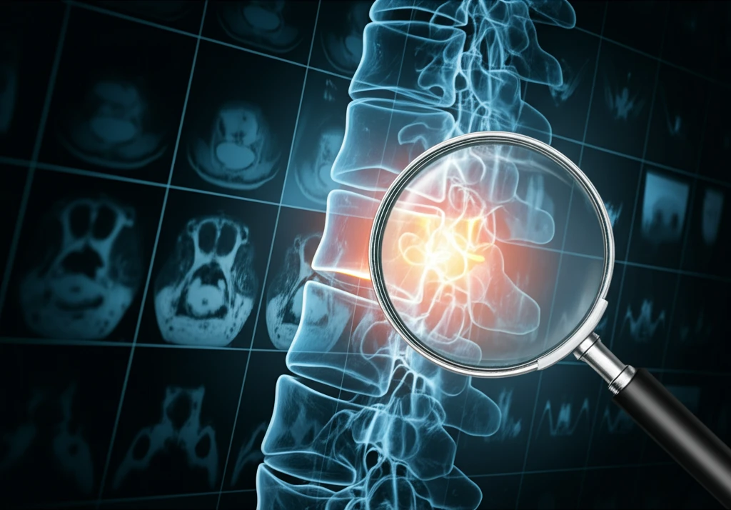
Decoding Back Pain: Is a Hidden Clue Lurking in Your MRI?
"Beyond the Usual Suspects: Exploring New MRI Findings in Spondyloarthritis"
Chronic back pain is a widespread issue, affecting millions and significantly impacting their quality of life. While many cases are attributed to common causes like muscle strain or disc problems, a subset stems from inflammatory conditions such as spondyloarthritis (SpA). Early diagnosis of SpA is crucial to manage the disease effectively and prevent long-term complications, yet it often presents a diagnostic challenge.
Magnetic resonance imaging (MRI) has become an indispensable tool in diagnosing various conditions, offering detailed insights into the body's internal structures. In the context of SpA, MRI helps visualize inflammation and structural changes in the sacroiliac joints and spine. However, traditional MRI assessments might overlook subtle yet significant signs that could indicate the presence of SpA, leading to delayed or missed diagnoses.
Emerging research suggests that specific MRI findings, particularly within the intervertebral spaces of the spine, could hold valuable clues for detecting SpA. These findings, which include unique signal intensities and structural changes, have the potential to enhance the accuracy of MRI in diagnosing SpA, offering new hope for individuals suffering from chronic back pain. The purpose of this article is to explore the cutting-edge approach and give you a better understanding of these underutilized markers and what they may mean for your health journey.
New Bone Formations: Spotting the Hidden Markers of Spondyloarthritis

A groundbreaking study has shed light on the significance of specific MRI findings in diagnosing spondyloarthritis (SpA), emphasizing the potential of new bone formations as key indicators. The study meticulously analyzed MRI scans of individuals with and without axial SpA, focusing on the intervertebral joints of the spine. The results revealed that certain signal intensities and structural changes, often overlooked in standard assessments, could serve as valuable diagnostic markers.
- Intradiscal High Signal Intensity: The presence of high signal intensity within the intervertebral disc, as seen on T1-weighted MRI images, was strongly associated with SpA.
- Vertebral Corner Bridging: The formation of bony bridges at the corners of the vertebrae was identified as a specific sign of SpA.
- Transdiscal Ankylosis: The fusion of vertebrae across the disc space was also found to be a significant marker.
Taking Control of Your Back Pain Journey
If you are experiencing chronic back pain, discussing these findings with your doctor can be a proactive step in your healthcare journey. While these MRI markers are highly specific, they may not be evident in every case of SpA. A comprehensive evaluation, including physical exams, medical history, and other diagnostic tests, is essential for an accurate diagnosis. By staying informed and engaged, you can work collaboratively with your healthcare team to explore all available options and find the most effective path toward relief and improved well-being.
