Uterus, Adenomyosis Diagnosis Through Ultrasound

Decoding Adenomyosis: How Ultrasound Can Offer Clarity

"A Deep Dive into the Accuracy of Transvaginal Ultrasound for Diagnosing Adenomyosis"


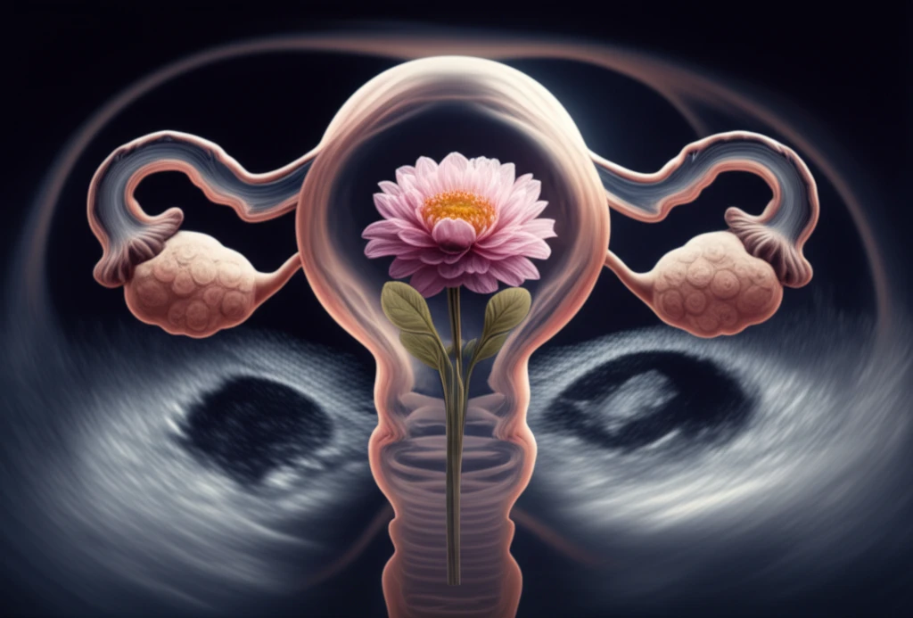
Adenomyosis, a condition where endometrial tissue grows into the muscular walls of the uterus, often brings a host of painful and disruptive symptoms. Diagnosing it has traditionally relied on post-surgical evaluations, but now, transvaginal ultrasound (TVUS) is stepping up as a non-invasive way to identify this condition early. This method offers a beacon of hope for those seeking answers without surgery.

Navigating the maze of adenomyosis diagnosis can be overwhelming. While TVUS has emerged as a promising tool, the lack of standardized imaging techniques and definitive markers has left many in the dark. This article aims to shed light on the accuracy of TVUS, focusing on its various features and how they contribute to a clearer diagnosis.

We'll explore how different imaging characteristics, such as 2D and 3D TVUS, elastography, and color Doppler, enhance diagnostic precision. By examining the latest research, this guide will help you understand the potential of TVUS in providing a non-surgical diagnosis, empowering you to make informed decisions about your health.

TVUS Unveiled: What the Studies Show

Uterus, Adenomyosis Diagnosis Through Ultrasound

A comprehensive review of recent studies highlights the capabilities of TVUS in diagnosing adenomyosis. The analysis included eight studies, evaluating various TVUS techniques and features. Let’s break down the key findings to understand how TVUS measures up.

When using 2D TVUS, the pooled sensitivity—its ability to correctly identify those with adenomyosis—was 83.8%, while its specificity—the ability to correctly identify those without the condition—was 63.9%. Certain imaging features stood out, particularly heterogeneous myometrium, which showed the highest sensitivity at 86.0%, and a globular uterus shape, which had the highest specificity at 78.1%. The 'question mark' sign, when combined with other TVUS features, notably boosted both sensitivity and specificity to 92% and 88%, respectively.

  • Sensitivity: Measures how well the test correctly identifies those who have the condition.
  • Specificity: Measures how well the test correctly identifies those who do not have the condition.
  • Heterogeneous Myometrium: Refers to the irregular appearance of the uterine muscle.
  • Globular Uterus: Describes an enlarged, rounded shape of the uterus.
Transitioning to 3D TVUS, the pooled sensitivity was 88.9%, with a specificity of 56.0%. While 3D imaging didn't significantly improve overall accuracy compared to 2D, it did offer unique insights. Poor definition of the junctional zone—the boundary between the endometrium and myometrium—showed the highest sensitivity at 86% and specificity at 56.0% with 3D TVUS. Preliminary data from TVUS with color Doppler indicated high sensitivity and specificity in differentiating between adenomyosis and myomas, while TVUS elastography showed enhanced specificity compared to 2D TVUS.

Empowering Diagnosis, Brighter Futures

The journey to diagnosing adenomyosis can be complex, but advancements in ultrasound technology provide clearer pathways. By understanding the strengths and limitations of TVUS, you can better navigate your diagnostic options. Keep in mind that while TVUS is a valuable tool, consulting with healthcare professionals remains crucial for personalized care. With ongoing research and improved imaging techniques, the future of adenomyosis diagnosis looks promising, offering hope for more accurate and timely interventions.

About this Article -

This article was crafted using a human-AI hybrid and collaborative approach. AI assisted our team with initial drafting, research insights, identifying key questions, and image generation. Our human editors guided topic selection, defined the angle, structured the content, ensured factual accuracy and relevance, refined the tone, and conducted thorough editing to deliver helpful, high-quality information.See our About page for more information.

Everything You Need To Know

1

What exactly is adenomyosis and why is it important to get diagnosed?

Adenomyosis is a condition where the endometrial tissue, which normally lines the uterus, grows into the muscular walls of the uterus. This can lead to painful and disruptive symptoms. It's important to diagnose this early to avoid long term pain. Early diagnosis has traditionally been challenging, often relying on post-surgical evaluations. However, Transvaginal Ultrasound (TVUS) is emerging as a non-invasive method to identify adenomyosis earlier.

2

What is Transvaginal Ultrasound (TVUS) and how does it help in diagnosing adenomyosis?

Transvaginal Ultrasound (TVUS) is a non-invasive imaging technique used to diagnose adenomyosis. During a TVUS, a probe is inserted into the vagina to visualize the uterus. This allows doctors to observe various characteristics and features that may indicate the presence of adenomyosis, such as a heterogeneous myometrium (irregular appearance of the uterine muscle) or a globular uterus (enlarged, rounded shape of the uterus). TVUS is significant because it offers a way to diagnose adenomyosis without surgery.

3

What do 'sensitivity' and 'specificity' mean in the context of TVUS results for adenomyosis?

Sensitivity measures the ability of a test, like Transvaginal Ultrasound (TVUS), to correctly identify individuals who have adenomyosis. Specificity, on the other hand, measures the ability of the test to correctly identify individuals who do not have adenomyosis. High sensitivity is crucial to avoid missing cases of adenomyosis, while high specificity is important to minimize false positives, ensuring that individuals without the condition are not misdiagnosed. For example, in studies involving 2D TVUS, heterogeneous myometrium showed high sensitivity, while a globular uterus shape had high specificity.

4

What is the difference between 2D and 3D TVUS, and which one is better for diagnosing adenomyosis?

2D TVUS and 3D TVUS are different approaches within Transvaginal Ultrasound. 2D TVUS provides two-dimensional images of the uterus. 3D TVUS offers three-dimensional views, potentially providing additional insights. While studies have shown that 3D TVUS doesn't significantly improve overall accuracy compared to 2D TVUS, it can highlight specific features like poor definition of the junctional zone (the boundary between the endometrium and myometrium). The choice between 2D and 3D TVUS often depends on the specific diagnostic goals and the expertise of the healthcare provider.

5

What are 'heterogeneous myometrium,' 'globular uterus,' and the 'question mark' sign, and how do they relate to adenomyosis diagnosis?

Heterogeneous myometrium refers to the irregular appearance of the uterine muscle as seen on Transvaginal Ultrasound (TVUS). This is a key imaging feature used in diagnosing adenomyosis. A globular uterus describes an enlarged, rounded shape of the uterus, which is another characteristic observed during TVUS. The 'question mark' sign is a specific visual pattern that, when combined with other TVUS features, can significantly improve the accuracy of adenomyosis diagnosis. These imaging characteristics help healthcare professionals to identify and diagnose adenomyosis more effectively.

Newsletter Subscribe

Subscribe to get the latest articles and insights directly in your inbox.