Cellular Communication: Unlocking Bioenergetics Through Membrane Contact Sites
"Scientists explore how organelles 'talk' to each other and why it matters for fighting disease."
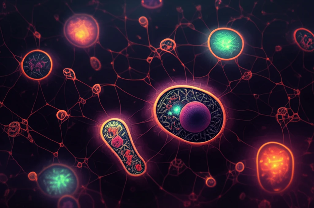
Imagine a bustling city where different departments work together seamlessly. Within our cells, tiny structures called organelles collaborate in a similar way. They connect at membrane contact sites (MCSs), which are like tiny bridges that allow the movement of molecules between them. These MCSs, typically closer than 30 nanometers, have been observed in yeast, plants, and even in us.
New research is investigating these cellular connections in Trypanosoma brucei, a parasite responsible for diseases like African trypanosomiasis (sleeping sickness). In mammalian cells, the endoplasmic reticulum (ER) connects with mitochondria to shuttle calcium ions (Ca2+), a process essential for energy production. The ER releases Ca2+ via inositol 1,4,5-trisphosphate receptors (InsP3R), facilitating mitochondrial uptake.
In trypanosomes, the InsP3R isn't in the ER; it's in acidocalcisomes—specialized storage compartments that act as the cell's main calcium reserves. This study uses advanced microscopy to find and describe the contact points between these acidocalcisomes and mitochondria, revealing how calcium is transferred. Understanding these interactions could unlock new ways to disrupt the parasite's energy and fight disease.
Why Understanding Organelle Communication is Key to New Therapies
The microscopic world within our cells is full of activity. Organelles, such as mitochondria and acidocalcisomes, do more than just exist in the same space—they physically interact. These points of contact, or membrane contact sites (MCSs), allow for direct communication through the exchange of ions and molecules.
- Visual Evidence: Using high-resolution microscopy, researchers confirmed direct contact between acidocalcisomes and mitochondria in T. brucei.
- Proximity Matters: Electron microscopy revealed that the membranes of these organelles were consistently less than 30 nanometers apart, allowing for quick molecule transfers.
- Life Stage Variations: Both the procyclic form (PCF) in insects and the bloodstream form (BSF) in mammals displayed these contact sites, suggesting their importance throughout the parasite's life cycle.
- Contact Site Confirmation: A proximity ligation assay (PLA) highlighted the close association between these organelles, marking them with fluorescent spots that could be easily observed.
Future Directions: Targeting MCSs for Therapeutic Intervention
By identifying and characterizing these membrane contact sites, researchers have opened a new avenue for developing targeted therapies against trypanosomiasis. Interfering with the formation or function of these MCSs could disrupt the parasite's ability to regulate calcium, produce energy, and ultimately survive within its host. This research lays the groundwork for innovative treatments that target the unique cellular biology of trypanosomes, offering hope for more effective and less toxic therapies.