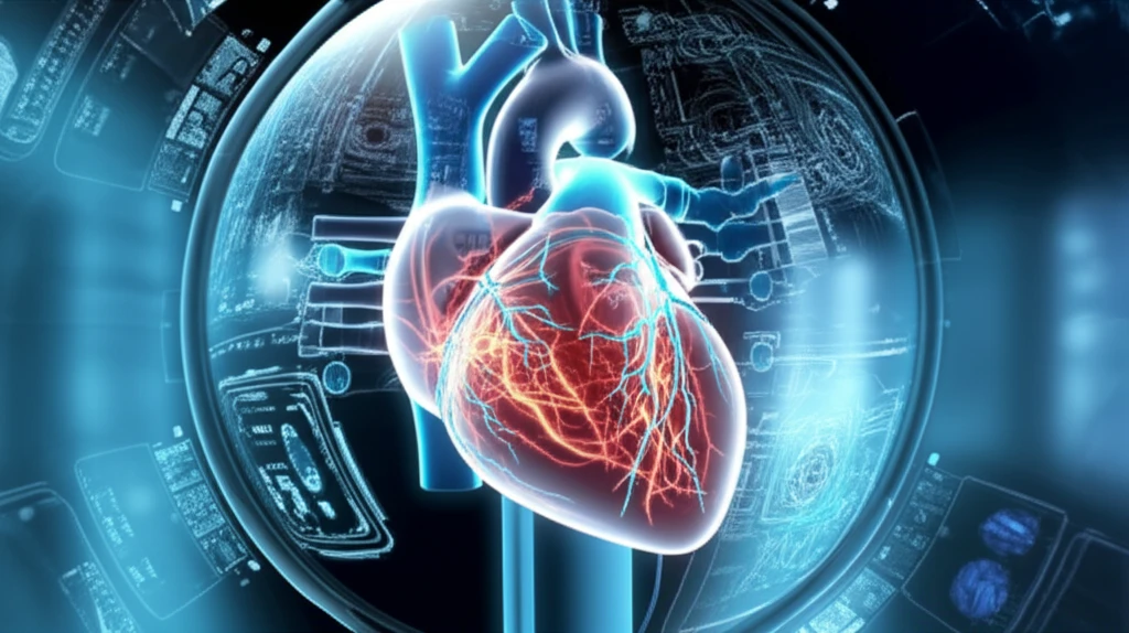
Heart undergoing repair with medical imaging technology

Cardiac Imaging: Revolutionizing Heart Attack Care

"Discover how advanced cardiac imaging is transforming the diagnosis and treatment of acute coronary syndromes, leading to better patient outcomes."


In the critical moments following a heart attack, timely and effective treatment is paramount. While initial emergency care focuses on rapid revascularization, the role of cardiac imaging becomes crucial in assessing the success of these interventions and understanding the long-term impact of ischemia and reperfusion on the heart.

Cardiac Magnetic Resonance (CMR) imaging stands out due to its unique ability to characterize myocardial tissue. CMR can detect early signs of myocardial edema, which appears soon after an ischemic event. These insights help differentiate between reversible and irreversible damage, guiding treatment strategies to salvage at-risk tissue.

This article delves into how cardiac imaging, particularly CMR, is transforming the management of acute coronary syndromes. We will explore how it aids in evaluating the effectiveness of revascularization, understanding the consequences of ischemia-reperfusion injury, and predicting long-term outcomes, ultimately leading to more personalized and effective patient care.

Unveiling the Acute Phase: How Imaging Spots Early Damage

Heart undergoing repair with medical imaging technology

During the initial diagnostic phase of an acute ST-elevation myocardial infarction (STEMI), imaging modalities have limited use, as immediate revascularization is the priority. However, imaging, especially CMR, shines in evaluating the effectiveness of revascularization and exploring the sequelae of prolonged ischemia and the consequences of reperfusion.

CMR's unique ability to explore the myocardium and characterize lesions allows for the early detection of myocardial edema, the first manifestation during the ischemic process, which can persist for up to 6 months post-event. These CMR studies have revealed that irreversible damage can occur as early as 90 minutes of myocardial ischemia, emphasizing the importance of rapid revascularization to save as much heart muscle as possible.

  • Early Edema Detection: CMR detects myocardial edema, a key early marker of ischemia.
  • Risk Area Identification: Differentiates between reversible edema and irreversible injury to define the myocardium at risk.
  • Prognostic Impact: Helps predict patient outcomes post-revascularization.
The extent of the 'myocardium at risk' significantly impacts patient prognosis after revascularization. Post-conditioning techniques have demonstrated effectiveness in limiting irreversible damage within this zone. CMR has enhanced our understanding of the stages of ischemia-reperfusion, guiding optimal management to minimize the effects of both ischemia and reperfusion. Despite advancements in treatment timing and revascularization techniques, some degree of infarction remains common in most STEMI cases. CMR helps quantify the extent of myocardial scarring and identifies complications like microvascular obstruction ('no reflow') and reperfusion hemorrhage, all of which significantly affect ventricular remodeling and arrhythmia risk.

Looking Ahead: Microcirculation and the Future of Cardiac Care

The ischemia-reperfusion process in acute coronary syndromes leads to early myocardial damage that can now be effectively assessed using CMR. Early detection of these lesions allows for evaluating the efficacy of revascularization and assessing myocardial prognosis.

The remodeling process that follows can be monitored by measuring strain in echocardiography. Future patient monitoring will likely incorporate coronary flow quantification techniques to measure coronary reserve, a marker of microvascular damage, which represents an initial step in the pathophysiology of ischemia-reperfusion.

By integrating advanced imaging techniques, particularly those targeting microvascular function, clinicians can gain a more comprehensive understanding of the individual patient's response to ischemia-reperfusion, paving the way for more tailored and effective treatment strategies. This will not only improve immediate outcomes but also reduce the long-term burden of heart disease.

About this Article -

This article was crafted using a human-AI hybrid and collaborative approach. AI assisted our team with initial drafting, research insights, identifying key questions, and image generation. Our human editors guided topic selection, defined the angle, structured the content, ensured factual accuracy and relevance, refined the tone, and conducted thorough editing to deliver helpful, high-quality information.See our About page for more information.

This article is based on research published under:

DOI-LINK: 10.1016/j.amcp.2018.06.010, Alternate LINK

Title: Ischémie–Reperfusion Dans Le Syndrome Coronaire Aigu St+ : Qu’Apporte L’Imagerie Cardiaque ?

Subject: Cardiology and Cardiovascular Medicine

Journal: Archives des Maladies du Coeur et des Vaisseaux - Pratique

Publisher: Elsevier BV

Authors: O. Lairez

Published: 2018-09-01

Everything You Need To Know

1

What is the primary role of cardiac imaging in the context of acute coronary syndromes?

Cardiac imaging is a pivotal tool in the diagnosis and treatment of acute coronary syndromes, especially after a heart attack. It focuses on assessing the effects of ischemia and reperfusion injuries. Its importance lies in guiding treatment strategies, evaluating the success of revascularization, and predicting patient outcomes. Specifically, Cardiac Magnetic Resonance (CMR) imaging is crucial in this context as it can characterize myocardial tissue and detect early signs of damage, like myocardial edema.

2

How does Cardiac Magnetic Resonance (CMR) imaging contribute to the management of heart attacks?

The role of Cardiac Magnetic Resonance (CMR) imaging is to characterize myocardial tissue, especially after a heart attack, to assess the impact of ischemia and reperfusion. During the initial phases of an acute ST-elevation myocardial infarction (STEMI), immediate revascularization is the primary focus. However, CMR excels in evaluating the efficacy of these interventions and in identifying the extent of myocardial damage. For instance, CMR can detect myocardial edema, which can help to identify if damage is reversible, or irreversible, and guide subsequent treatment.

3

What is the significance of myocardial edema, as detected by Cardiac Magnetic Resonance (CMR), in assessing heart damage?

Myocardial edema, as detected by Cardiac Magnetic Resonance (CMR) imaging, is a significant early marker of ischemia. It appears soon after an ischemic event and can persist for up to six months. Early detection of myocardial edema allows clinicians to differentiate between reversible and irreversible damage. This distinction is critical because it directly influences treatment decisions aimed at salvaging at-risk myocardial tissue and improving patient prognosis. Understanding the extent of myocardial edema is key to assessing the effectiveness of revascularization.

4

How does Cardiac Magnetic Resonance (CMR) imaging help in evaluating the success of revascularization techniques?

Revascularization techniques aim to restore blood flow to the heart muscle after a heart attack. Cardiac Magnetic Resonance (CMR) imaging plays a vital role in evaluating the success of these techniques. By assessing the extent of myocardial damage and the presence of complications like microvascular obstruction and reperfusion hemorrhage, CMR helps in determining if revascularization has been successful in minimizing irreversible injury. It also helps in understanding the long-term impact of these interventions on patient outcomes and future treatment strategies.

5

In what ways does cardiac imaging, particularly Cardiac Magnetic Resonance (CMR), influence patient outcomes?

The use of Cardiac Magnetic Resonance (CMR) imaging affects patient outcomes by guiding treatment strategies and predicting long-term prognosis. CMR helps in assessing the extent of myocardial damage, identifying complications like microvascular obstruction, and evaluating the effectiveness of revascularization. This information allows for more personalized and effective patient care, leading to better outcomes and a more accurate assessment of the risk of ventricular remodeling and arrhythmia. Understanding the stages of ischemia-reperfusion helps in optimizing patient management.

Newsletter Subscribe

Subscribe to get the latest articles and insights directly in your inbox.