Microscopic view of glowing cancer cells being scanned in blood vessels.

Can a New Blood Test Spot Cancer Earlier? The Promise of 'Liquid Biopsies'

"Researchers are developing innovative methods to detect and analyze circulating tumor cells, potentially revolutionizing cancer diagnostics and treatment monitoring."


For decades, the fight against cancer has centered on early detection. The sooner cancer is identified, the better the chances of successful treatment and survival. While traditional methods like imaging and biopsies have been crucial, they often detect cancer at later stages. Now, a groundbreaking approach is emerging that promises to revolutionize cancer diagnostics: the "liquid biopsy."

Liquid biopsies involve analyzing circulating tumor cells (CTCs) in the bloodstream. These cells, shed from primary tumors, hold vital clues about the cancer’s characteristics and stage. Detecting and analyzing CTCs could provide a non-invasive way to monitor cancer progression, assess treatment effectiveness, and even predict recurrence.

Recent advancements in in vivo flow cytometry are significantly enhancing the capabilities of liquid biopsies. This sophisticated technique allows real-time identification and enumeration of apoptotic (dying) CTCs directly in the blood flow, offering a more dynamic and comprehensive understanding of the disease.

The Science Behind the Breakthrough: How In Vivo Flow Cytometry Works

Microscopic view of glowing cancer cells being scanned in blood vessels.

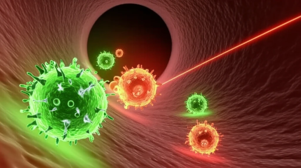
In vivo flow cytometry represents a significant leap forward in CTC detection. Unlike traditional methods that require drawing blood and analyzing it in a lab, this technique allows for real-time monitoring of cells directly within the bloodstream. Here’s a simplified breakdown:

  • Real-Time Monitoring: A laser irradiates circulating objects in the blood flow. This prompts any fluorescent-labeled cells to emit light, which is then detected by a highly sensitive photodetector.

  • Targeted Labeling: Researchers use fluorescent tags that specifically bind to cancer cells or markers of apoptosis (cell death). In this study, they used green fluorescent protein (GFP) to identify breast cancer cells (MDA-MB-231) and Annexin-V-RPE, an orange dye, to tag cells undergoing apoptosis.
  • Simultaneous Detection: The system can detect multiple fluorescent signals simultaneously, allowing for the differentiation between viable CTCs and apoptotic CTCs. This is crucial because only viable CTCs can create distant metastases.
  • High Sensitivity: The technique is highly sensitive, capable of detecting even small numbers of CTCs amidst the vast background of blood cells and tissue. This is vital for early detection and monitoring treatment response.
This method circumvents many limitations of traditional liquid biopsies. By analyzing cells in their natural environment, it minimizes the risk of altering cell properties or inducing artificial apoptosis, which can occur during sample preparation. It also provides a more accurate snapshot of the dynamic changes happening within the tumor microenvironment.

Future Implications: The Path to Personalized Cancer Care

The development of advanced in vivo flow cytometry techniques holds immense promise for the future of cancer treatment. By providing a real-time, non-invasive method to monitor CTCs and their apoptotic state, this technology can enable more personalized and effective cancer therapies. It can also help oncologists quickly assess treatment response, adjust treatment plans as needed, and ultimately improve patient outcomes. The ability to detect and analyze CTCs in real-time opens new avenues for understanding cancer metastasis and developing targeted therapies to prevent it.

About this Article -

This article was crafted using a human-AI hybrid and collaborative approach. AI assisted our team with initial drafting, research insights, identifying key questions, and image generation. Our human editors guided topic selection, defined the angle, structured the content, ensured factual accuracy and relevance, refined the tone, and conducted thorough editing to deliver helpful, high-quality information.See our About page for more information.

Everything You Need To Know

1

What is a liquid biopsy and how does it differ from traditional cancer detection methods?

A liquid biopsy is a non-invasive method that analyzes circulating tumor cells (CTCs) found in the bloodstream. Unlike traditional methods like imaging and biopsies, which often detect cancer at later stages, liquid biopsies offer the potential for earlier detection. By analyzing CTCs, which are shed from primary tumors, liquid biopsies provide vital clues about the cancer's characteristics, stage, and progression. This approach minimizes the invasiveness associated with traditional biopsies and allows for more frequent monitoring to assess treatment effectiveness and predict recurrence. The use of in vivo flow cytometry further enhances liquid biopsies by enabling real-time analysis of CTCs directly in the blood flow, a significant advancement over methods that require blood to be drawn and processed in a lab.

2

How does in vivo flow cytometry contribute to the advancements in cancer diagnostics?

In vivo flow cytometry represents a significant leap forward in cancer diagnostics by allowing real-time identification and enumeration of CTCs directly within the bloodstream. This technique uses a laser to irradiate circulating objects in the blood flow, prompting fluorescent-labeled cells to emit light. Researchers use fluorescent tags, such as green fluorescent protein (GFP) to identify breast cancer cells (MDA-MB-231), and Annexin-V-RPE, an orange dye, to tag cells undergoing apoptosis. The system's ability to simultaneously detect multiple fluorescent signals allows for differentiation between viable CTCs and apoptotic CTCs. This provides a more dynamic and comprehensive understanding of the disease, critical for early detection and monitoring treatment response.

3

Can you explain the process of how in vivo flow cytometry identifies and analyzes circulating tumor cells?

The process of in vivo flow cytometry involves several key steps. First, a laser irradiates the blood flow, and any fluorescent-labeled cells emit light. Researchers use targeted labeling with fluorescent tags, such as GFP to identify MDA-MB-231 cells and Annexin-V-RPE for apoptosis. The system's high sensitivity enables detection of even small numbers of CTCs. This technique allows for real-time monitoring within the bloodstream, minimizing the risk of altering cell properties or inducing artificial apoptosis, unlike traditional methods. It provides a more accurate snapshot of the dynamic changes within the tumor microenvironment. The detection of both viable and apoptotic CTCs is crucial because only viable CTCs can create distant metastases. This provides a more comprehensive understanding of the cancer's progression.

4

What are the advantages of in vivo flow cytometry compared to traditional liquid biopsy techniques?

In vivo flow cytometry offers several advantages over traditional liquid biopsy techniques. By analyzing cells in their natural environment, in vivo flow cytometry minimizes the risk of altering cell properties or inducing artificial apoptosis, a common concern with traditional methods. It provides a more accurate snapshot of the dynamic changes happening within the tumor microenvironment. Additionally, this technique enables real-time monitoring, which is not possible with traditional methods that require blood to be drawn and analyzed in a lab. The high sensitivity of in vivo flow cytometry is also a significant benefit, allowing for the detection of even small numbers of CTCs, which is critical for early detection and monitoring treatment response.

5

How might the development of in vivo flow cytometry techniques impact the future of cancer treatment and patient care?

The development of advanced in vivo flow cytometry techniques holds immense promise for the future of cancer treatment. By providing a real-time, non-invasive method to monitor CTCs and their apoptotic state, this technology can enable more personalized and effective cancer therapies. Oncologists can use this technology to quickly assess treatment response, adjust treatment plans as needed, and ultimately improve patient outcomes. The ability to detect and analyze CTCs in real-time opens new avenues for understanding cancer metastasis and developing targeted therapies to prevent it. This will likely lead to earlier detection, more tailored treatments, and better overall survival rates for cancer patients.

Newsletter Subscribe

Subscribe to get the latest articles and insights directly in your inbox.