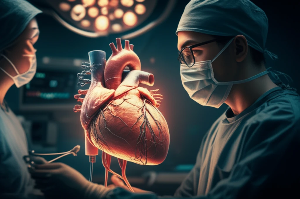
CABG Check-Up: Ensuring Graft Success During Coronary Bypass
"A guide to the latest techniques in intraoperative graft assessment, ensuring optimal outcomes in coronary artery bypass surgery."
Coronary artery bypass graft (CABG) surgery has become a cornerstone in treating coronary artery disease since its introduction in the 1960s. Its long-term success depends significantly on graft patency—how well the new grafts stay open and functional. Early graft failure, especially within the first year, can often be traced back to technical errors during the surgery itself.
That's why assessing these grafts right in the operating room is so vital. This allows surgeons to identify and correct any issues immediately, securing the graft's function and, ultimately, improving patient outcomes. But how exactly do surgeons evaluate these delicate new connections during the procedure?
This article explores the latest techniques for intraoperative graft assessment, focusing on transit-time flowmetry (TTFM) and fluorescence imaging. We'll break down how these methods work, their benefits, limitations, and what they mean for the future of CABG surgery. Understanding these techniques can empower you to ask informed questions and advocate for the best possible care.
TTFM: Measuring Blood Flow in Real-Time
Transit-time flowmetry (TTFM) is the most common method used during surgery to assess graft function. It uses ultrasound technology to measure blood flow in the newly implanted grafts.
- Mean Graft Flow (MGF): The average blood flow through the graft, measured in milliliters per minute (mL/min).
- Pulsatility Index (PI): Indicates the resistance to blood flow within the graft and the downstream coronary arteries.
- Backward Flow Percentage (%BF): Measures the amount of blood flowing backward through the graft during the cardiac cycle.
- Diastolic Filling Percentage (DF%): Represents the proportion of blood flow occurring during the heart's relaxation phase (diastole).
The Future of Graft Assessment: A Combined Approach
While TTFM is a valuable tool, it has limitations. Factors like blood pressure and the condition of the downstream coronary arteries can influence TTFM readings. Newer techniques like high-resolution epicardial ultrasonography (HR-ECUS) can be used alongside TTFM to provide a more complete picture. HR-ECUS uses ultrasound to visualize the graft and surrounding vessels, helping surgeons pinpoint the exact location of any blockages or narrowing.
Fluorescence imaging (IFI) is another promising technology. It involves injecting a special dye that lights up under infrared light, allowing surgeons to see the blood flow in the graft in real-time. While IFI shows great promise, more research is needed to fully understand its role in CABG surgery.
The ongoing development and refinement of these intraoperative assessment techniques hold the key to improving the long-term success of CABG surgery. By combining these advanced tools, surgeons can make informed decisions, optimize graft function, and ultimately improve the lives of patients with coronary artery disease.