Mycobacterium ulcerans, the bacterium responsible for Buruli ulcer, glowing blue bacterium in wetlands, affecting skin and health.

Buruli Ulcer: Unlocking the Secrets of a Neglected Disease

"A Deep Dive into Genotyping Tools, Transmission Mysteries, and Future Prospects for Combating Mycobacterium ulcerans"


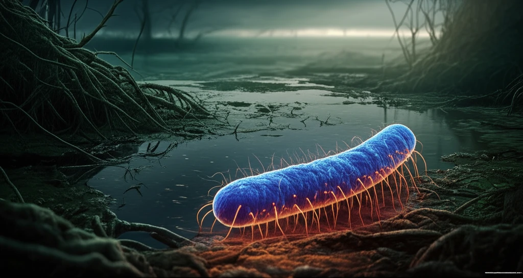
Buruli ulcer, caused by the environmental bacterium Mycobacterium ulcerans (MU), is a debilitating skin and soft tissue infection affecting people in over 30 countries. Though treatable, it remains a significant public health challenge due to its elusive transmission routes and the lack of precise diagnostic tools, especially in resource-limited settings. Understanding how this infection spreads and developing better ways to identify and track it are critical for effective prevention and control.

Current research efforts are heavily focused on deciphering the ecology of M. ulcerans, particularly its presence and behavior in aquatic environments, and pinpointing the exact mechanisms by which it transmits to humans. Since M. ulcerans is difficult to culture from environmental samples, scientists rely on molecular methods, especially genotyping, to differentiate it from other similar bacteria and track its spread. Genotyping allows researchers to compare strains from different sources, identify potential reservoirs, and potentially trace the source of outbreaks.

This article will explore the genotyping tools currently available for M. ulcerans, focusing on their strengths, weaknesses, and potential for improvement. Furthermore, we'll discuss how these tools, coupled with advancements in other fields, can enhance our understanding of M. ulcerans transmission, evolution, and the emergence of drug resistance. By improving diagnostic methods and refining genotyping tools, we can better complement public health efforts to reduce the burden of Buruli ulcer.

Decoding M. ulcerans: The Role of Genotyping

Mycobacterium ulcerans, the bacterium responsible for Buruli ulcer, glowing blue bacterium in wetlands, affecting skin and health.

Genotyping tools serve as powerful methods to distinguish M. ulcerans strains from other Mycolactone-Producing Mycobacteria (MPMs). This differentiation is crucial because these bacteria share similar characteristics but may have different implications for human health. Key tools include:

Beyond simply identifying the presence of M. ulcerans, genotyping can help us understand the micro-geographical distribution of different strains. M. ulcerans thrives in aquatic environments, particularly slow-moving water bodies and wetlands with specific pH ranges. Understanding these ecological niches can help us target interventions and minimize human contact with contaminated water sources. Studies have found higher concentrations of M. ulcerans DNA in biofilms compared to other water samples, indicating that efforts to culture the bacteria from environmental sources should focus on methods that concentrate biofilms.

  • IS2404 and IS2606: These insertion sequences are genetic markers used to differentiate MPMs from M. marinum.
  • Enoyl Reductase (ER) and Keto Reductase (KR) genes: These genes, involved in mycolactone synthesis, are also utilized to detect and differentiate between M. ulcerans, M. liflandii, and M. marinum DL.
  • Variable Number Tandem Repeats (VNTR): VNTR analysis has been used to resolve genetic homogeneity within and between geographical isolates, differentiating M. ulcerans from other MPMs.
Early and accurate diagnosis is essential for effective Buruli ulcer treatment. Traditional methods like microscopy for acid-fast bacilli (AFB) have limitations in sensitivity and specificity. The WHO recommends supplementary tests like culture, PCR, or histopathology for confirming cases. While histopathology can reveal necrosis in soft tissues, it doesn't pinpoint M. ulcerans specifically. Culture methods are time-consuming, taking weeks to yield visible colonies. PCR and real-time PCR offer rapid and accurate diagnosis, but their high cost limits their accessibility in resource-poor areas. Loop-Mediated Isothermal Amplification (LAMP) has emerged as a cost-effective alternative, showing superiority to PCR in some studies and making it a promising tool for point-of-care diagnosis.

Charting the Course: The Future of M. ulcerans Research

As cases of Buruli ulcer continue to surface, refining our understanding of M. ulcerans remains paramount. Integrating molecular biology, genomics, and bioinformatics will accelerate progress in understanding the mechanisms of infection, pathology, and treatment. Rescanning published genomes and refining existing tools can unveil nuanced sequences to differentiate MU strains, while integrating VNTR and SNP analysis can reveal strains becoming resistant. Ultimately, cumulative efforts in improving diagnostic methods and fine-tuning genotyping tools are essential for elucidating transmission routes, studying the molecular epidemiology of MU, and detecting emerging resistance.

About this Article -

This article was crafted using a human-AI hybrid and collaborative approach. AI assisted our team with initial drafting, research insights, identifying key questions, and image generation. Our human editors guided topic selection, defined the angle, structured the content, ensured factual accuracy and relevance, refined the tone, and conducted thorough editing to deliver helpful, high-quality information.See our About page for more information.

Everything You Need To Know

1

What is Buruli ulcer and what are the challenges in combating it?

Buruli ulcer is a debilitating skin and soft tissue infection caused by the environmental bacterium Mycobacterium ulcerans (MU). The disease poses a significant public health challenge due to its elusive transmission routes and the lack of precise diagnostic tools, especially in resource-limited settings. These factors hinder effective prevention and control efforts, necessitating a deeper understanding of how MU spreads and the development of improved diagnostic methods.

2

How do genotyping tools aid in understanding and combating Mycobacterium ulcerans?

Genotyping tools are crucial for differentiating Mycobacterium ulcerans (MU) strains from other Mycolactone-Producing Mycobacteria (MPMs). Key tools include IS2404 and IS2606, which are genetic markers used to differentiate MPMs from M. marinum, Enoyl Reductase (ER) and Keto Reductase (KR) genes involved in mycolactone synthesis, and Variable Number Tandem Repeats (VNTR). These tools allow researchers to track the spread of MU, identify potential reservoirs, and potentially trace the source of outbreaks, enhancing our understanding of its micro-geographical distribution and ecological niches.

3

What are the current diagnostic methods for Buruli ulcer and their limitations?

Current diagnostic methods include microscopy for acid-fast bacilli (AFB), culture, PCR, real-time PCR, and histopathology. Microscopy has limitations in sensitivity and specificity. Culture methods are time-consuming, taking weeks to yield visible colonies. PCR and real-time PCR offer rapid and accurate diagnosis but are costly. Histopathology reveals necrosis but doesn't pinpoint Mycobacterium ulcerans specifically. Loop-Mediated Isothermal Amplification (LAMP) has emerged as a cost-effective alternative, showing superiority to PCR in some studies.

4

How does the environment influence the spread of Mycobacterium ulcerans?

Mycobacterium ulcerans (MU) thrives in aquatic environments, particularly slow-moving water bodies and wetlands with specific pH ranges. Studies have found higher concentrations of MU DNA in biofilms compared to other water samples. Understanding these ecological niches is vital for targeting interventions and minimizing human contact with contaminated water sources. This ecological understanding helps in predicting potential outbreak areas and designing prevention strategies.

5

What are the future prospects for Mycobacterium ulcerans research and how can we improve control of Buruli ulcer?

The future of Mycobacterium ulcerans (MU) research involves integrating molecular biology, genomics, and bioinformatics to understand infection mechanisms, pathology, and treatment. Refining existing tools and integrating VNTR and SNP analysis can reveal strains becoming resistant. Improving diagnostic methods and fine-tuning genotyping tools are essential for elucidating transmission routes, studying the molecular epidemiology of MU, and detecting emerging resistance. The combined effort of advanced research, refined tools, and better understanding of the bacterium will improve the control of Buruli ulcer.

Newsletter Subscribe

Subscribe to get the latest articles and insights directly in your inbox.