Illustration of opposing morphogen gradients forming an embryo.

Building Blocks of Life: How Scientists Created a Vertebrate Embryo From Scratch

"Revolutionary research unveils the minimum requirements for embryonic development, paving the way for regenerative medicine."


The development of a vertebrate embryo is a marvel of biological engineering, orchestrated by complex molecular and cellular processes. These processes progressively instruct embryonic cells, assigning them their identities and dictating their behaviors. Over the past two decades, researchers have identified numerous genes crucial to vertebrate embryo development [1, 2].

However, a fundamental question remained unanswered: What are the minimal conditions required to instruct pluripotent embryonic cells and organize them into distinct tissues and organs? This challenge was taken up, seeking the experimental conditions necessary to construct a complete embryo by instructing early-stage zebrafish (Danio rerio) cells using a combination of morphogenic signals.

The quest to understand how embryos are formed has a rich history. Nearly a century ago, Hilde Mangold, a student in Hans Spemann's lab, conducted a pivotal experiment. She transplanted the dorsal blastopore lip from a gastrula of one species of newt (Triturus taeniatus) to the ventral side of a gastrula of another species (Triturus cristatus, whose cells are unpigmented) [3]. This resulted in the formation of a secondary embryonic axis at the transplantation site.

The Organizer Concept and Its Limitations: Spemann's Legacy

Illustration of opposing morphogen gradients forming an embryo.

The secondary axis comprised pigmented cells from the donor (Triturus taeniatus) and unpigmented cells from the host (Triturus cristatus). This remarkable ability to organize host cells led to the dorsal blastopore lip being defined as an 'organizer' and subsequently known as the 'Spemann organizer.' Over the past two decades, the molecular nature of this dorsal organizer has been elucidated [4].

Key to its activity are factors secreted by the dorsal blastopore lip, acting as antagonists of ventral morphogens and establishing a ventro-dorsal activity gradient (Figure 1A). In Spemann and Mangold's pioneering experiment, dorsal factors secreted by the graft inhibited ventral morphogen activity at the transplantation site. This generated a new gradient, mirroring the morphogenic activity gradient in the wild-type embryo's lateral and dorsal parts (Figure 1B).

  • However, when this organizer is grafted into a neutral environment, such as the animal pole of a zebrafish blastula, its activity is limited.
  • Only axial mesendodermal tissues like the notochord form [5].
  • This result suggested that the organizing activity extends beyond this dorsal center.
Further experiments involving grafting different portions of the zebrafish blastula or gastrula margin to the animal pole of a host blastula revealed that the ventral embryonic margin acts as a caudal organizer [5]. In contrast, the lateral and dorso-lateral regions of the embryonic margin organize the trunk and posterior cephalic region, respectively [6]. Therefore, contrary to the prevailing dogma, the organizing activities are distributed along the embryonic margin, forming a continuum from the dorsal to the ventral territory [6].

Minimum Requirements for an Embryo: Two Gradients

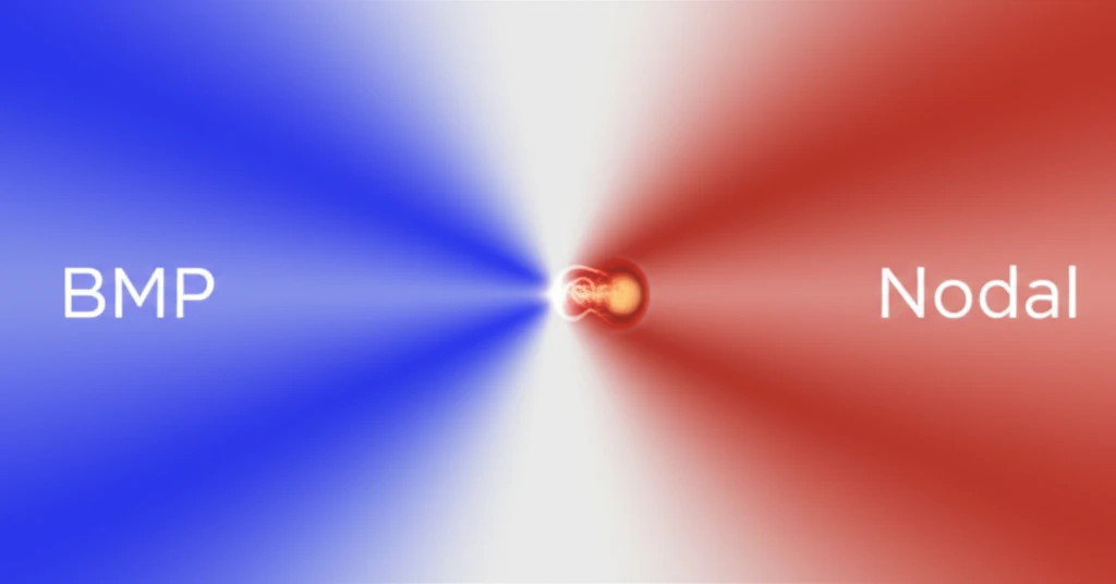
The embryonic margin expresses numerous inducing factors, notably Nodal and BMP (bone morphogenetic protein). The BMP signaling pathway is active ventrally, decreasing towards the dorsal margin. Conversely, Nodal activity is present in all marginal cells, with higher concentration dorsally (Figure 1C). Thus, there’s continuous variation in the BMP/Nodal activity ratio from ventral regions where it’s high, to dorsal regions where it’s negligible (Figure 1C). Opposing gradients of Nodal and BMP define the minimal conditions for organizing a complete embryonic axis.

About this Article -

This article was crafted using a human-AI hybrid and collaborative approach. AI assisted our team with initial drafting, research insights, identifying key questions, and image generation. Our human editors guided topic selection, defined the angle, structured the content, ensured factual accuracy and relevance, refined the tone, and conducted thorough editing to deliver helpful, high-quality information.See our About page for more information.

Everything You Need To Know

1

How does a vertebrate embryo actually form and why is understanding this important?

A vertebrate embryo is created through complex molecular and cellular processes that instruct embryonic cells, assigning them identities and dictating their behaviors. This organized development is essential for forming distinct tissues and organs. Understanding this process can unlock advancements in regenerative medicine, enabling the repair or replacement of damaged tissues.

2

What is the Spemann organizer and why was it such an important discovery?

The Spemann organizer, initially identified as the dorsal blastopore lip, has the remarkable ability to induce the formation of a secondary embryonic axis when transplanted. This is significant because it demonstrates the power of specific embryonic regions to organize surrounding cells. However, its activity is limited to forming axial mesendodermal tissues like the notochord, indicating the existence of other organizing centers within the embryo.

3

What are morphogen gradients and why are they essential for embryonic development?

Morphogen gradients are concentration gradients of signaling molecules that provide positional information to cells during development. Specifically, opposing gradients of Nodal and BMP (bone morphogenetic protein) define the minimal conditions for organizing a complete embryonic axis. These gradients dictate cell fate and differentiation, ensuring the proper spatial arrangement of tissues and organs. Disruptions in morphogen gradients can lead to developmental abnormalities.

4

Besides the dorsal blastopore lip, what other regions of the embryo have organizing activities?

The ventral embryonic margin acts as a caudal organizer, and the lateral and dorso-lateral regions organize the trunk and posterior cephalic region, respectively. This challenges the idea that the Spemann organizer is the sole organizing center, instead suggesting a continuum of organizing activities distributed along the embryonic margin. Understanding the functions of these different regions is crucial for a complete picture of embryonic development.

5

How do Nodal and BMP contribute to the formation of an embryo?

Nodal is present in all marginal cells, with higher concentrations dorsally, while BMP signaling is active ventrally, decreasing towards the dorsal margin. The ratio between these two morphogens dictates cell fate and tissue organization. This balance ensures that the embryo develops properly along its axes, with appropriate structures forming in the correct locations.

Newsletter Subscribe

Subscribe to get the latest articles and insights directly in your inbox.