Glowing skeletal system of fetus inside womb.

Building Blocks: How Fetal Bone Development Sets the Stage for Lifelong Health

"Unlocking the secrets of bone formation in the womb to understand and improve bone health across the lifespan. Understanding human bone Development is the first step."


Our comprehension of human bone development, notably during the prenatal period, has undergone significant advancements, primarily fueled by the study of animal models. Direct investigations into human bone development remain essential, however, due to the crucial insights that pathological and genetic discoveries from human bone disorders provide. These findings not only give rise to new research questions but also validate observations from model organisms, revealing previously unknown mechanisms in bone formation.

Moreover, it's important to recognize that animal models cannot fully replicate all aspects of human conditions. This article concentrates on human-derived data pertaining to the physiology of bone development in fetuses and newborns. It also addresses intrinsic and extrinsic elements influencing bone health, offering a comprehensive view of how these factors contribute to both typical and abnormal bone development.

We will also discuss the impact that maternal nutrition, environmental factors, and genetics play in shaping the skeletal health of future generations.

What are the Stages of Fetal and Neonatal Bone Development?

Glowing skeletal system of fetus inside womb.

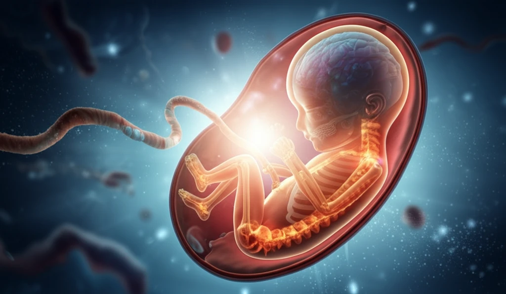
The skeletal system's pattern is largely set by the start of fetal development, which occurs eight weeks following fertilization. Newborns are about 12 times longer in body length than the earliest fetus (30mm versus 360mm in crown-rump length). Rapid growth is thus the primary theme of bone development during the fetal period. For example, between 16 and 41 weeks of gestation, the femur elongates at a rate of 0.35 mm per day [1]. Bone formation, or ossification, is a vital process in bone development and growth. It requires the coordination of vasculogenesis, mineralization, matrix production, and osteoblast differentiation.

Studies have indicated that the majority of bones start to ossify in the first few weeks of the fetal stage, and that ossification centers appear sequentially in each bone. The ossification of the clavicle, humerus, and mandible, for example, takes place during the embryonic stage (6–7 weeks). The ossification of the talus or cuboid, in contrast, doesn't start until late in pregnancy, around week 28, or after birth [2].

  • Clavicle, humerus, and mandible ossify during the embryonic stage (6-7 weeks).
  • Talus or cuboid ossification starts late, around week 28, or after birth.
Secondary ossification centers (SOCs) form in the cartilage epiphysis of the long bones, beginning with the creation of cartilage canals. These canals form when perichondrium invades the epiphysis center. This mechanism introduces mesenchymal cells and vasculature into the epiphysis, which promotes bone formation [3]. Vascular invasion and the start of cartilage canal development in the epiphysis of the distal femur begin between weeks 8 and 10 of pregnancy. A complex vascular system is fully formed within the canals by week 14, several months before SOC development [4]. Except for the clavicles, where SOCs do not appear until 18 to 20 years of age [5], most SOCs appear between the late embryonic stage and a few years after birth.

Optimizing Bone Health From the Start

Understanding the complex processes of fetal and neonatal bone development provides a foundation for promoting lifelong bone health. By focusing on maternal nutrition, reducing exposure to harmful environmental factors, and addressing genetic predispositions, healthcare providers and parents can work together to ensure that every child has the best possible start on the path to strong, healthy bones. Early intervention and continued monitoring are key to addressing any developmental issues and maximizing long-term skeletal well-being.

About this Article -

This article was crafted using a human-AI hybrid and collaborative approach. AI assisted our team with initial drafting, research insights, identifying key questions, and image generation. Our human editors guided topic selection, defined the angle, structured the content, ensured factual accuracy and relevance, refined the tone, and conducted thorough editing to deliver helpful, high-quality information.See our About page for more information.

This article is based on research published under:

DOI-LINK: 10.1002/9781119266594.ch15, Alternate LINK

Title: Human Fetal And Neonatal Bone Development

Journal: Primer on the Metabolic Bone Diseases and Disorders of Mineral Metabolism

Publisher: Wiley

Authors: Tao Yang, Monica Grover, Kyu Sang Joeng, Brendan Lee

Published: 2018-09-28

Everything You Need To Know

1

What key processes are essential for bone formation, and when does ossification typically begin for different bones during fetal development?

During fetal bone development, the skeletal system's pattern is largely established by the eighth week following fertilization. At this stage, rapid growth is the primary characteristic. Bone formation, also known as ossification, is crucial, requiring coordinated vasculogenesis, mineralization, matrix production, and osteoblast differentiation. Ossification centers appear sequentially in each bone, with bones like the clavicle, humerus, and mandible ossifying earlier (6-7 weeks) compared to bones like the talus or cuboid (around week 28 or after birth).

2

How do secondary ossification centers (SOCs) form, and what role do cartilage canals play in this process within fetal long bones?

Secondary ossification centers (SOCs) develop in the cartilage epiphysis of long bones, initiating with the creation of cartilage canals. These canals form when the perichondrium invades the epiphysis center, introducing mesenchymal cells and vasculature, which promotes bone formation. Vascular invasion and the start of cartilage canal development in the epiphysis of the distal femur begin between weeks 8 and 10 of pregnancy. Most SOCs appear between the late embryonic stage and a few years after birth, except for the clavicles, where SOCs do not appear until 18 to 20 years of age.

3

What are the primary factors that influence fetal bone development, and how can healthcare providers and parents ensure a child's optimal skeletal health from the start?

Maternal nutrition, environmental factors, and genetics significantly influence fetal bone development. Maternal health directly impacts a child's skeletal future. Optimizing bone health from the start involves focusing on maternal nutrition, minimizing exposure to harmful environmental factors, and addressing genetic predispositions. Healthcare providers and parents play a crucial role in ensuring a child's best possible start toward strong, healthy bones through early intervention and continued monitoring.

4

What specific elements must be coordinated during ossification, and while exercise during fetal development isn't directly discussed, how might fetal positioning relate to bone development?

The process of ossification requires the coordination of several key elements: vasculogenesis, which establishes the necessary blood supply; mineralization, which provides the essential minerals for bone hardness; matrix production, which forms the structural framework of the bone; and osteoblast differentiation, where specialized cells create new bone. Problems with any of these coordinated activities can cause developmental issues. While the impact of exercise or mechanical loading during fetal stages isn't directly addressed, it's understood that mechanical stimuli generally support bone development postnatally. Thus, ensuring proper fetal positioning and movement may indirectly support healthy bone development.

5

How has the study of animal models advanced our understanding of fetal bone development, and why is human-derived data still essential in this field?

Animal models have greatly contributed to our understanding of human bone development, especially during prenatal stages. However, direct investigations into human bone development are crucial due to unique insights from pathological and genetic discoveries related to human bone disorders. These findings validate observations from model organisms and uncover previously unknown mechanisms in bone formation. Because animal models cannot fully replicate all aspects of human conditions, focusing on human-derived data provides a more accurate view of fetal and newborn bone development.

Newsletter Subscribe

Subscribe to get the latest articles and insights directly in your inbox.