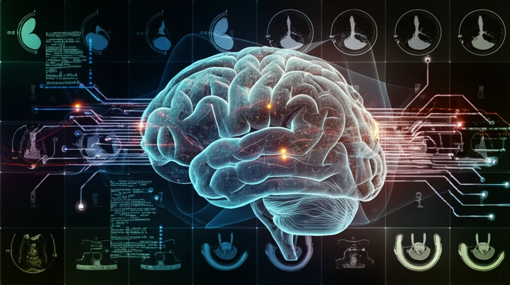
AI-Enhanced Brain Tumor Analysis

Brain Tumor Breakthrough: AI-Powered Analysis for Accurate Diagnosis

"Revolutionizing Glioma Grading with 18F-FET-PET and Perfusion MRI Texture Features"


Gliomas, which constitute a significant 80% of all malignant brain tumors, present a considerable challenge in oncology. Accurate classification of these tumors is critical for surgeons and oncologists to formulate effective treatment plans. This is where advanced imaging techniques and artificial intelligence come into play, promising a new era of precision in diagnostics.

Perfusion-weighted MRI (PWI) stands out as an advanced magnetic resonance imaging technique, offering quantifiable hemodynamic parameters that include relative cerebral blood volume (rCBV), cerebral blood flow (rCBF), and mean transit time (MTT). Positron emission tomography (PET) with 18F-FET further aids in delineating cerebral gliomas and differentiating between high- and low-grade tumors. The fusion of these technologies holds potential for significantly improving diagnostic accuracy.

This analysis delves into a study evaluating the use of 18F-FET-PET and perfusion-weighted MRI texture features. By adopting a principal component analysis (PCA), the study aims to refine the classification of high- and low-grade gliomas. PCA helps to reduce the number of features needed, addressing the common challenge of limited patient data in this specialized area of research.

Decoding the Study: How AI Analyzes Brain Tumor Images

AI-Enhanced Brain Tumor Analysis

The study, conducted between 2012 and 2016, involved 27 patients diagnosed with brain tumors—18 with high-grade gliomas and 9 with low-grade gliomas. These patients underwent both FET-PET and perfusion MRI imaging. The MRI imaging was performed using a Siemens 3T Tim Trio Scanner, part of a hybrid 3TMR-BrainPET setup. Dynamic susceptibility-weighted contrast-enhanced T2-MRI acquisition was performed during the first pass of a contrast agent, using echo-planar imaging (EPI).

Following the imaging process, perfusion maps were generated from the MRI images to quantify cerebral hemodynamics. These maps, along with the FET-PET images, were co-registered using PMOD (PMOD Ltd.) image fusion tools, which apply automatic matching methods based on rigid registration. Tumor volumes of interest (VOIs) were identified in the FET-PET images using a threshold-based method, with the minimum uptake threshold set at 1.6 times the average background region. Background regions were defined as spheres of 15 mm³.

Here’s a breakdown of the key steps in image processing:
  • Image Co-registration: Aligning PET and MRI images for accurate comparison.
  • VOI Creation: Delineating tumor regions based on FET-PET uptake.
  • Normalization: Standardizing perfusion maps using PMOD software.
  • Texture Feature Extraction: Analyzing gray-level patterns within VOIs using various matrices.
To extract meaningful information from the images, several texture feature matrices were employed, including the gray level co-occurrence matrix (GLCM), the neighborhood gray-level different matrix (NGLDM), the gray level run length matrix (GLRLM), and the gray level zone length matrix (GLZLM), alongside conventional histogram features. This comprehensive analysis was performed individually for each VOI derived from FET-PET and normalized perfusion maps.

The Future of Brain Tumor Diagnostics

This study illustrates how texture feature analysis, combined with PCA, can significantly reduce the number of features required for 18F-FET PET and MRI data analysis. By reducing the feature set to one-fourth of the original size, PCA streamlines the analytical process and enhances the focus on the most critical data. This approach not only makes the analysis more manageable but also sets the stage for more accurate and efficient diagnostic models. As data collection expands and analytical techniques advance, the potential for creating predictive models capable of classifying gliomas with greater precision becomes increasingly viable, heralding a new era in personalized cancer treatment.

About this Article -

This article was crafted using a human-AI hybrid and collaborative approach. AI assisted our team with initial drafting, research insights, identifying key questions, and image generation. Our human editors guided topic selection, defined the angle, structured the content, ensured factual accuracy and relevance, refined the tone, and conducted thorough editing to deliver helpful, high-quality information.See our About page for more information.

Everything You Need To Know

1

Why is accurate glioma grading so crucial, and how does artificial intelligence support it?

Gliomas, accounting for 80% of malignant brain tumors, pose significant challenges in oncology. Artificial intelligence, specifically texture analysis on medical imaging like Perfusion-weighted MRI and 18F-FET-PET scans, aids in accurate glioma grading. Correct classification is crucial because it allows surgeons and oncologists to create better treatment plans, ultimately improving patient outcomes.

2

What imaging techniques, such as 18F-FET-PET and perfusion MRI, were used in the brain tumor study, and what specific equipment was involved?

The study utilized both 18F-FET-PET and perfusion MRI. The MRI scans were conducted using a Siemens 3T Tim Trio Scanner. During the scans, dynamic susceptibility-weighted contrast-enhanced T2*-MRI acquisition was performed during the first pass of a contrast agent, using echo-planar imaging (EPI). These imaging techniques provide complementary information about the tumor's metabolic activity and hemodynamic characteristics.

3

Can you break down the key steps in image processing the AI uses to analyze brain tumor images?

The study leverages several key image processing steps: Image Co-registration aligns PET and MRI images, VOI Creation delineates tumor regions based on 18F-FET-PET uptake, Normalization standardizes perfusion maps using PMOD software, and Texture Feature Extraction analyzes gray-level patterns within VOIs using various matrices. These steps help extract meaningful, quantifiable data from the medical images.

4

How does Principal Component Analysis (PCA) help in streamlining the data analysis for 18F-FET PET and MRI?

The study reduced the feature set to one-fourth of its original size through Principal Component Analysis (PCA). This streamlined the analytical process and focuses analysis on the most critical data derived from 18F-FET PET and MRI. By diminishing the number of features needed, PCA addresses the challenge of limited patient data, making the analysis more manageable and accurate.

5

What are the future implications of using AI-powered texture analysis and PCA for brain tumor diagnostics and personalized treatment?

By combining texture feature analysis with PCA, the classification of gliomas can become more precise. Future research could involve larger datasets and more advanced analytical techniques that can enhance the accuracy of predictive models. These models may enable personalized cancer treatments, marking a significant advancement in brain tumor diagnostics and patient care. The potential for using AI to tailor treatments based on individual tumor characteristics is a promising avenue for future exploration.

Newsletter Subscribe

Subscribe to get the latest articles and insights directly in your inbox.