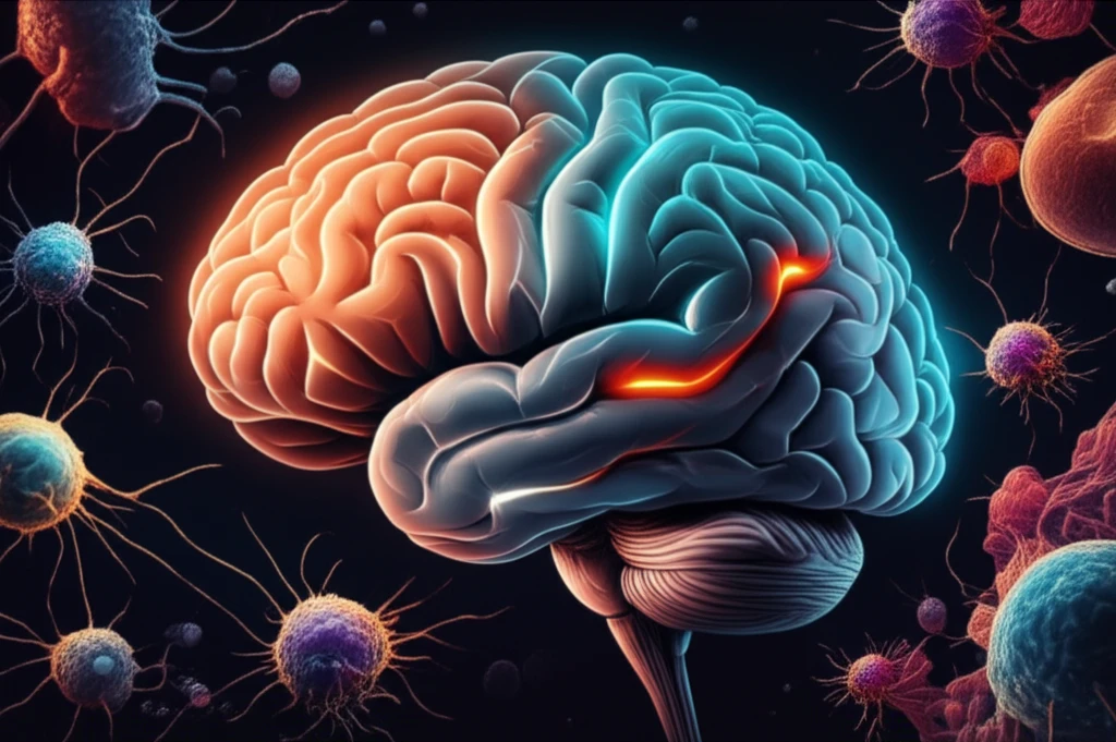
MRI scan highlighting areas of brain damage.

Brain Injury Breakthrough: MRI Scans Expose Hidden Damage

"New research reveals how advanced MRI can detect subtle changes in the brain after an injury, offering hope for better diagnosis and treatment."


Traumatic brain injury (TBI) affects millions globally, leading to disability and death. While the immediate effects of a TBI can be obvious, the long-term consequences and subtle brain changes are often missed by standard imaging techniques. This gap in diagnostic capability can delay appropriate treatment and hinder recovery.

Traditionally, doctors have relied on CT scans and conventional MRI to assess brain damage. However, these methods often fail to detect microstructural changes in the brain's gray matter – the area responsible for higher-level cognitive functions. As a result, many TBI patients may appear to have 'normal' scans despite experiencing debilitating symptoms.

Now, a groundbreaking study is shedding light on how advanced MRI techniques, particularly Diffusion Tensor Imaging (DTI) and Diffusion Kurtosis Imaging (DKI), can reveal these hidden injuries. These methods offer a more detailed picture of the brain's microstructural integrity, opening new avenues for diagnosis and targeted therapies.

Unveiling Hidden Damage with Advanced MRI

MRI scan highlighting areas of brain damage.

Researchers utilized advanced MRI techniques – Diffusion Tensor Imaging (DTI) and Diffusion Kurtosis Imaging (DKI) – to visualize subtle changes in the gray matter of rodent brains following controlled cortical impact (CCI), a model for TBI. They followed these changes over a month, with MRI scans performed at 5 hours, 1, 3, 7, 14, and 30 days post-injury.

To confirm the MRI findings, the researchers also examined the brain tissue under a microscope, using cresyl violet staining and Glial Fibrillary Acidic Protein (GFAP) immunohistochemistry. This approach allowed them to link changes observed on MRI with actual tissue damage and cellular responses.

  • DTI: Measures the direction and magnitude of water diffusion in the brain, revealing information about white matter integrity.
  • DKI: An extension of DTI that models non-Gaussian water diffusion, providing more detailed information about tissue microstructure and complexity.
  • GFAP: An indicator of astrogliosis, the proliferation of astrocytes (a type of glial cell) in response to injury.
The results demonstrated that DTI and DKI could detect changes in the brain's gray matter following injury, including increases in fractional anisotropy (FA) in the early stages after injury, followed by decreases in FA in later stages. Also decreased mean diffusivity (MD) in the injured area, indicative of cytotoxic edema or cell swelling. The study also revealed changes in the contralateral hemisphere, the side of the brain opposite the injury, suggesting that TBI can have widespread effects.

Future Implications for TBI Treatment

This study provides valuable insights into the dynamic changes that occur in the brain after a TBI, and highlights the potential of advanced MRI techniques to improve diagnosis, monitor disease progression, and develop targeted therapies. Further research is needed to translate these findings into clinical practice, with the ultimate goal of improving outcomes for individuals affected by TBI.

About this Article -

This article was crafted using a human-AI hybrid and collaborative approach. AI assisted our team with initial drafting, research insights, identifying key questions, and image generation. Our human editors guided topic selection, defined the angle, structured the content, ensured factual accuracy and relevance, refined the tone, and conducted thorough editing to deliver helpful, high-quality information.See our About page for more information.

Everything You Need To Know

1

What is Traumatic Brain Injury (TBI) and why is it a significant health concern?

Traumatic Brain Injury (TBI) is a serious condition affecting millions globally, often leading to long-term disability or even death. The impact of a TBI can range widely, from mild concussions to severe injuries. The primary concern stems from the potential for delayed diagnosis due to the limitations of standard imaging techniques, which can lead to delayed or inadequate treatment, impacting patient recovery and quality of life. Understanding the complexities of TBI and having access to advanced diagnostic tools are crucial for improving patient outcomes and advancing neurological care.

2

How do traditional imaging techniques like CT scans and conventional MRI fall short in diagnosing TBI?

Traditional methods such as CT scans and conventional MRI often struggle to detect subtle changes in the brain's gray matter following a Traumatic Brain Injury (TBI). These methods are primarily designed to identify large-scale damage, such as bleeding or swelling, but they often miss microstructural alterations. Because these changes are not readily visible, many individuals with TBI may be misdiagnosed or face delays in receiving appropriate treatment, which can significantly affect their long-term recovery. The study highlights the importance of more sensitive imaging techniques to uncover these hidden injuries and improve patient care.

3

What are Diffusion Tensor Imaging (DTI) and Diffusion Kurtosis Imaging (DKI), and how do they improve TBI diagnosis?

Diffusion Tensor Imaging (DTI) and Diffusion Kurtosis Imaging (DKI) are advanced MRI techniques used to visualize subtle brain changes after a Traumatic Brain Injury (TBI). DTI measures the direction and magnitude of water diffusion within the brain, providing insights into white matter integrity. DKI, which builds upon DTI, provides even more detailed information about tissue microstructure and complexity. These methods enable researchers to detect microstructural changes in the brain's gray matter, which is often missed by conventional imaging. This detailed information aids in better diagnosis and can inform the development of targeted therapies by revealing hidden injuries that were previously undetectable.

4

What were the key findings when using advanced MRI techniques, such as DTI and DKI, in the study of TBI?

The study utilized Diffusion Tensor Imaging (DTI) and Diffusion Kurtosis Imaging (DKI) to reveal important changes in the brain after a Traumatic Brain Injury (TBI). The research showed that DTI and DKI could detect changes in the gray matter. The findings included alterations in fractional anisotropy (FA), with increases in early stages followed by decreases later. Also, the researchers observed a decrease in mean diffusivity (MD) in the injured area, signaling cytotoxic edema. Additionally, the study highlighted that the effects of TBI are not confined to the injury site, revealing changes in the contralateral hemisphere, indicating widespread damage.

5

How might the use of advanced MRI techniques like DTI and DKI impact the future treatment of TBI?

The future of Traumatic Brain Injury (TBI) treatment looks promising with the use of advanced MRI techniques like Diffusion Tensor Imaging (DTI) and Diffusion Kurtosis Imaging (DKI). These techniques offer a more detailed view of brain damage, allowing for earlier and more accurate diagnoses. This could enable clinicians to monitor disease progression with greater precision, leading to the development of more targeted therapies. Further research is crucial to translate these findings into clinical practice, with the ultimate goal of improving outcomes for individuals affected by TBI by improving diagnosis, monitoring, and treatment strategies. This can involve personalized treatment plans and more effective interventions to improve patient recovery and quality of life.

Newsletter Subscribe

Subscribe to get the latest articles and insights directly in your inbox.