Illustration of a brain scan with MRI technology showing brain injury detection

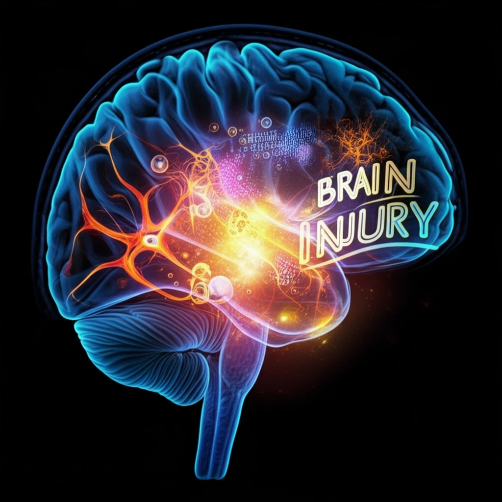
Brain Injury Breakthrough: How MRI is Unveiling Hidden Damage & Paving the Way for Better Treatment

"Groundbreaking research reveals that advanced MRI techniques can detect subtle brain changes after injury, offering new hope for diagnosis and recovery."


Brain injuries, whether from a car accident, a sports injury, or a fall, can have devastating and lasting effects. For years, doctors have relied on traditional methods like CT scans and standard MRIs to assess the damage. However, these methods often miss the subtle changes that occur at the microscopic level, leaving a crucial part of the injury's story untold.

Now, a new study published in the Journal of Neurotrauma is shedding light on this hidden world. Researchers are using advanced MRI techniques to reveal the intricate ways brain tissue changes after an injury. This breakthrough offers a more complete picture of the damage and paves the way for more effective treatments.

This article will delve into the innovative MRI methods used, what the researchers discovered about the brain's response to injury, and the potential this has for the future of brain injury diagnosis and care. We'll explore how this research could change the lives of millions who experience these types of injuries each year.

Unveiling the Hidden: How Advanced MRI Works

Illustration of a brain scan with MRI technology showing brain injury detection

The study used cutting-edge MRI methods to look beyond the basics and examine what happens to the brain after an injury. These methods are known as Diffusion Tensor Imaging (DTI) and Diffusion Kurtosis Imaging (DKI).

These advanced techniques allow researchers to examine how water molecules move through the brain's tissue. The way water moves can reveal important information about the health and structure of the brain's cells and fibers. DTI and DKI give doctors a much more detailed view of the damage.

  • DTI (Diffusion Tensor Imaging): This technique tracks the movement of water molecules along the brain's fibers, which are responsible for communication. Changes in this movement can indicate damage to these crucial pathways.
  • DKI (Diffusion Kurtosis Imaging): Building on DTI, DKI provides even more detail about the complexity of water diffusion, helping to identify subtle changes in brain tissue structure, like swelling or inflammation.
By using these methods, researchers can now see the changes happening at the microscopic level. This level of insight is essential for understanding the full impact of a brain injury.

A Brighter Future for Brain Injury Care

This research marks a significant step forward in understanding and treating brain injuries. By using advanced MRI, doctors can gain a deeper insight into the damage, paving the way for more effective diagnosis, treatment, and, ultimately, better outcomes for those affected by brain injuries. This will enable more effective and targeted therapies, offering hope to millions affected by this type of injury.

About this Article -

This article was crafted using a human-AI hybrid and collaborative approach. AI assisted our team with initial drafting, research insights, identifying key questions, and image generation. Our human editors guided topic selection, defined the angle, structured the content, ensured factual accuracy and relevance, refined the tone, and conducted thorough editing to deliver helpful, high-quality information.See our About page for more information.

Everything You Need To Know

1

What are the limitations of traditional methods like CT scans and standard MRIs in assessing brain injuries?

Traditional methods like CT scans and standard MRIs often miss subtle changes that occur at the microscopic level in the brain tissue after an injury. These methods may not reveal the intricate details of damage to the brain's cells and fibers, leaving a crucial part of the injury's story untold. Advanced MRI techniques such as Diffusion Tensor Imaging (DTI) and Diffusion Kurtosis Imaging (DKI) are necessary to get a complete picture.

2

How do advanced MRI techniques like Diffusion Tensor Imaging (DTI) and Diffusion Kurtosis Imaging (DKI) work to reveal hidden brain damage?

Diffusion Tensor Imaging (DTI) tracks the movement of water molecules along the brain's fibers, which are responsible for communication. Changes in this movement can indicate damage to these crucial pathways. Diffusion Kurtosis Imaging (DKI) builds on DTI by providing even more detail about the complexity of water diffusion, helping to identify subtle changes in brain tissue structure, such as swelling or inflammation. These methods allow researchers to see changes happening at the microscopic level, providing essential insights into the full impact of a brain injury.

3

What is the significance of using Diffusion Tensor Imaging (DTI) and Diffusion Kurtosis Imaging (DKI) in understanding brain injuries?

The significance of using Diffusion Tensor Imaging (DTI) and Diffusion Kurtosis Imaging (DKI) lies in their ability to reveal the hidden, microscopic changes in the brain that traditional methods miss. By tracking water molecule movement, DTI can detect damage to the brain's communication fibers. DKI provides even greater detail, identifying subtle changes in tissue structure like swelling or inflammation. This level of insight is crucial for a comprehensive understanding of the injury's impact and for developing targeted treatments.

4

How might the use of advanced MRI techniques, such as Diffusion Tensor Imaging (DTI) and Diffusion Kurtosis Imaging (DKI), improve treatment outcomes for brain injuries?

By using advanced MRI techniques like Diffusion Tensor Imaging (DTI) and Diffusion Kurtosis Imaging (DKI), doctors can gain a deeper insight into the specific types and locations of brain damage. This detailed understanding paves the way for more effective and targeted therapies, potentially leading to better outcomes for those affected by brain injuries. This could involve tailored rehabilitation programs, pharmaceutical interventions, or other treatments designed to address the specific microscopic damages identified by the advanced imaging.

5

What implications does the increased use of Diffusion Tensor Imaging (DTI) and Diffusion Kurtosis Imaging (DKI) have on the future of brain injury diagnosis and care?

The increased use of Diffusion Tensor Imaging (DTI) and Diffusion Kurtosis Imaging (DKI) signifies a move towards more precise and personalized brain injury diagnosis and care. With the ability to detect subtle, microscopic changes in brain tissue, doctors can develop more targeted and effective treatment plans. This could lead to improved rehabilitation strategies, pharmaceutical interventions, and a better understanding of the long-term effects of brain injuries. Moreover, this advanced imaging can aid in tracking the progress of recovery and adjusting treatment accordingly, offering hope for millions affected by this type of injury.

Newsletter Subscribe

Subscribe to get the latest articles and insights directly in your inbox.