Abstract representation of brain connectivity using ICA and ROI methods.

Brain Graphs Unveiled: How Data-Driven Methods Are Revolutionizing Neuroscience

"Discover how Independent Component Analysis (ICA) is reshaping our understanding of brain connectivity, outperforming traditional ROI methods in fMRI analysis."


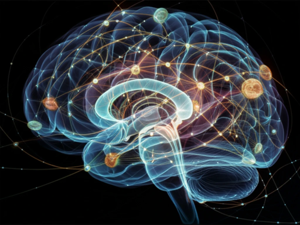
The human brain, a complex network of interconnected regions, has always been a subject of intense scientific curiosity. Modern neuroscience relies heavily on functional Magnetic Resonance Imaging (fMRI) to map and understand the intricate relationships between different brain areas. One powerful approach to analyzing fMRI data is graph theory, which allows researchers to quantitatively characterize the architecture of these brain networks.

Graph theory-based analysis involves representing the brain as a graph, where nodes represent brain regions and edges represent the connections between them. A key challenge is defining these nodes. Two popular methods have emerged: defining nodes based on regions of interest (ROIs) determined by brain atlases, or using spatial brain components estimated by independent components analysis (ICA).

A groundbreaking study has shed light on the effectiveness of these two methods. By using simulated fMRI data, researchers were able to compare the accuracy of graph metrics derived from ICA and ROI-based approaches. The results have significant implications for how neuroscientists construct and interpret brain graphs.

ICA vs. ROI: Why Data-Driven Approaches Are Gaining Ground

Abstract representation of brain connectivity using ICA and ROI methods.

Traditionally, ROIs defined by brain atlases have been a common choice for defining nodes in brain graphs. However, this approach has limitations. Brain atlases are based on anatomical structures, which may not perfectly align with functional boundaries. This can lead to the inclusion of functionally heterogeneous voxels within a single ROI, potentially skewing the results of the network analysis.

ICA, on the other hand, is a data-driven technique that identifies spatially independent components in the brain. These components represent functional networks that are naturally present in the data. This approach offers several advantages:

  • Functional Homogeneity: ICA components tend to be more functionally homogeneous than ROIs defined by anatomical atlases.
  • Spatial Relationships: ICA can capture complex spatial relationships between brain regions that may not be apparent from anatomical data alone.
  • Reduced Artifacts: ICA can help to mitigate the influence of artifacts in the data, leading to more accurate network representations.
The study compared graph metrics derived from ICA and ROI-based approaches to a known ground truth in simulated fMRI data. The results consistently showed that ICA-based graphs were more accurate than ROI-based graphs. This suggests that ICA is a more reliable method for defining nodes in brain graphs, particularly when studying complex brain networks.

Implications for Future Research

This research has significant implications for future studies of brain connectivity. By demonstrating the superiority of ICA-based methods, the study encourages researchers to adopt data-driven approaches for defining nodes in brain graphs. This can lead to more accurate and reliable characterizations of brain networks in both healthy individuals and those with neurological or psychiatric disorders. As neuroscience continues to evolve, embracing advanced analytical techniques like ICA will be crucial for unraveling the mysteries of the human brain.

About this Article -

This article was crafted using a human-AI hybrid and collaborative approach. AI assisted our team with initial drafting, research insights, identifying key questions, and image generation. Our human editors guided topic selection, defined the angle, structured the content, ensured factual accuracy and relevance, refined the tone, and conducted thorough editing to deliver helpful, high-quality information.See our About page for more information.

Everything You Need To Know

1

What is Independent Component Analysis (ICA), and why is it important in understanding brain connectivity?

Independent Component Analysis (ICA) is a data-driven technique used to identify spatially independent components in the brain from fMRI data. These components represent functional networks naturally present in the data, allowing researchers to map brain connectivity more effectively than traditional ROI methods. ICA's significance lies in its ability to capture functional homogeneity, complex spatial relationships, and reduce artifacts, leading to more accurate network representations. The implication is a shift towards data-driven approaches in neuroscience for better understanding of brain networks.

2

What are brain graphs, and why is their construction significant in neuroscience?

Brain graphs are constructed by representing brain regions as nodes and the connections between them as edges. This allows researchers to quantitatively characterize the architecture of brain networks derived from fMRI data. It's important because it provides a framework for understanding how different brain areas interact and communicate. The implications of brain graph construction are far-reaching, as they enable the study of brain connectivity in both healthy individuals and those with neurological disorders, potentially leading to new insights into brain function and disease.

3

What are Regions of Interest (ROIs), and what are the limitations of using them in brain network analysis?

Regions of Interest (ROIs) are brain regions defined by brain atlases and traditionally used as nodes in brain graphs. However, this approach has limitations because brain atlases are based on anatomical structures, which may not perfectly align with functional boundaries. This can lead to the inclusion of functionally heterogeneous voxels within a single ROI, potentially skewing the results of network analysis. While ROIs provide a structured way to analyze brain regions, the implications of their use must be carefully considered, especially when compared to data-driven approaches like ICA.

4

What is functional Magnetic Resonance Imaging (fMRI), and why is it important for studying the brain?

Functional Magnetic Resonance Imaging (fMRI) is a neuroimaging technique used to map and understand the intricate relationships between different brain areas. It works by detecting changes in blood flow in the brain, which are correlated with neural activity. fMRI is important because it allows researchers to non-invasively study brain function in both healthy individuals and those with neurological or psychiatric disorders. The implications of fMRI extend to various fields, including neuroscience, psychology, and medicine, as it provides valuable insights into brain function and dysfunction.

5

Why are data-driven approaches becoming more popular in neuroscience compared to traditional methods?

Data-driven approaches, such as ICA, are gaining ground in neuroscience due to their ability to overcome the limitations of traditional ROI-based methods. Unlike ROIs, which are based on anatomical structures, data-driven approaches identify functional networks directly from fMRI data. This leads to more accurate and reliable characterizations of brain networks, as they are not constrained by predefined anatomical boundaries. The implications of this shift towards data-driven approaches are significant, as they pave the way for a deeper understanding of brain connectivity and its role in various cognitive processes and neurological disorders.

Newsletter Subscribe

Subscribe to get the latest articles and insights directly in your inbox.