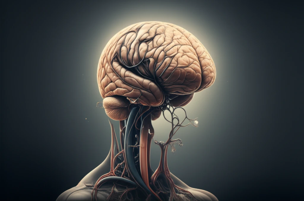
Brain Bleed or Contrast Leak? A Rare Post-Angiography Mystery
"When a routine heart procedure leads to a brain scare: understanding the complexities of diagnosis and potential risks."
In the realm of cardiac care, coronary angiography (CAG) stands as a crucial diagnostic tool, allowing physicians to visualize the heart's blood vessels and identify potential blockages. While generally safe, CAG, like any invasive procedure, carries inherent risks. Bleeding, a common complication of antithrombotic treatments in acute coronary syndrome (ACS), can be exacerbated by invasive procedures.
Contrast media (CM) and heparin, essential components of CAG, contribute to this risk profile. While the overall bleeding rate in ACS patients undergoing percutaneous coronary intervention (PCI) is significant, a far less common but potentially devastating complication is subarachnoid hemorrhage (SAH) – bleeding in the space surrounding the brain.
This article delves into a fascinating and rare case where a patient undergoing CAG developed SAH, mimicking contrast media leakage. It highlights the diagnostic challenges, the importance of considering alternative explanations, and the need for vigilance in post-CAG monitoring.
The Case: A Diagnostic Puzzle

A 55-year-old man with a history of a previous heart attack (acute ST elevation myocardial infarction or STEMI) was admitted for a follow-up CAG. The procedure itself was seemingly routine: a transradial approach, a minimal dose of heparin for catheter lubrication, and a standard amount of non-ionic contrast media (iodixanol) for visualization.
- Initial Suspicion: Contrast media leakage due to the immediate timing and the CT scan findings.
- Further Investigation: Cerebral angiography was performed to rule out vascular malformations.
- The Twist: No malformations were found, and the patient showed partial recovery, only to reveal subacute SAH on a follow-up MRI two weeks later.
Key Takeaways: Vigilance and Comprehensive Diagnosis
This case highlights the importance of maintaining a high index of suspicion for SAH following CAG, even in the absence of obvious risk factors or with seemingly clear initial explanations. While contrast media-induced neurotoxicity is a recognized phenomenon, clinicians must be vigilant in considering other potential causes of bleeding.
The use of advanced imaging techniques like MRI, in addition to CT scans, can be invaluable in differentiating between contrast leakage and true SAH, especially when symptoms are atypical or evolve over time. Furthermore, a thorough neurological assessment and continuous monitoring are essential in the post-CAG period.
Ultimately, this case serves as a reminder that in medicine, the most accurate diagnosis often requires a comprehensive approach, considering all possibilities and utilizing the full range of available diagnostic tools.
