Comparison of thick and thin-slice CT scans in a trauma setting

Beyond the Scan: Unveiling the Truth About Trauma CT and Slice Thickness

"Does a Thinner Slice Always Mean a Better Diagnosis? Exploring the Impact of CT Scan Technology in Trauma Cases"


In the fast-paced world of medical imaging, the quest for sharper, more detailed views is relentless. For trauma patients, where every second counts, this quest takes on even greater significance. Computed Tomography (CT) scans have become indispensable in quickly identifying internal injuries, guiding life-saving interventions. But a key question lingers: does the thickness of the CT scan slices truly affect the accuracy of diagnosis, especially in critical trauma situations?

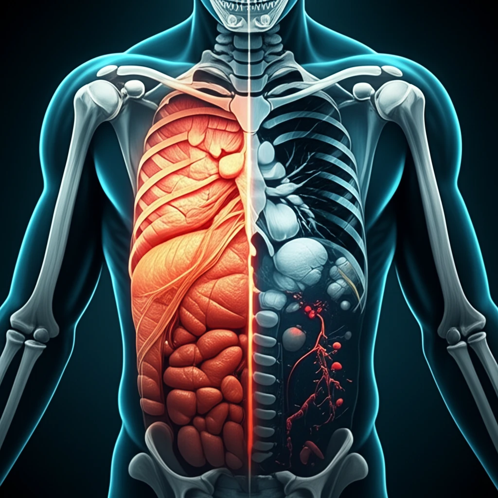
This article delves into a specific aspect of this technology: the comparison of thick-slice (5 mm) and thin-slice (1.5 mm) images in thoracoabdominal trauma CT scans. We will explore how these different scanning techniques influence the detection of injuries in the chest and abdomen, looking at soft tissue, bones, and lung structures. The aim is to provide a clear understanding of the benefits and limitations of each approach, offering insights for both healthcare professionals and those curious about the latest developments in medical imaging.

The study which this article is based on, compared the effectiveness of thick- and thin-slice CT scans in identifying injuries. The findings may surprise you, challenging some common assumptions about the impact of slice thickness on diagnostic accuracy and patient care.

Decoding Trauma CT: Why Slice Thickness Matters

Comparison of thick and thin-slice CT scans in a trauma setting

CT scans utilize X-rays to create detailed cross-sectional images of the body, enabling doctors to visualize internal organs and structures without invasive procedures. In trauma cases, the speed and accuracy of CT scans are crucial. They help in the detection of life-threatening injuries, like internal bleeding, organ damage, and fractures.

The concept of 'slice thickness' refers to the width of the images created by the CT scanner. Historically, thicker slices were common, but advances in technology have made thinner slices possible. Thinner slices offer higher resolution, potentially revealing more detail, but they also come with tradeoffs, such as increased scan time and higher radiation exposure.

  • Thick Slices: Typically, 5 mm or wider. They are faster to acquire and process, making them suitable for quickly identifying major injuries.
  • Thin Slices: Often 1.5 mm or narrower. They provide more detailed images, which can be helpful in detecting subtle injuries or smaller structures.
  • The Trade-off: While thinner slices offer more detail, they can also increase the scan time and radiation dose. This can impact how efficiently patients are managed in an emergency situation.
The original research aimed to compare thick (5 mm) and thin (1.5 mm) slice images in thoracoabdominal trauma CT. The study focused on detecting injuries in the lung, soft tissue, and bone windows of the scans, including specific injuries such as pneumothorax, organ damage, and fractures. The researchers also analyzed reconstruction and evaluation times to assess the practical implications of each technique.

Conclusion: Balancing Detail and Efficiency in Trauma Care

In the realm of trauma care, the choice between thick and thin slices in CT scans involves a delicate balance. The research underscores that while thinner slices can offer more detail, they do not necessarily translate into a significant improvement in detecting lung and soft tissue injuries. The study highlights the importance of considering the entire clinical picture, with the goal of providing the best possible care in the most efficient manner. As technology continues to evolve, the focus remains on optimizing imaging techniques to enhance diagnostic accuracy while prioritizing patient safety and well-being.

About this Article -

This article was crafted using a human-AI hybrid and collaborative approach. AI assisted our team with initial drafting, research insights, identifying key questions, and image generation. Our human editors guided topic selection, defined the angle, structured the content, ensured factual accuracy and relevance, refined the tone, and conducted thorough editing to deliver helpful, high-quality information.See our About page for more information.

This article is based on research published under:

DOI-LINK: 10.1007/s00068-018-1021-9, Alternate LINK

Title: Comparison Of Thick- And Thin-Slice Images In Thoracoabdominal Trauma Ct: A Retrospective Analysis

Subject: Critical Care and Intensive Care Medicine

Journal: European Journal of Trauma and Emergency Surgery

Publisher: Springer Science and Business Media LLC

Authors: Leon Guchlerner, Julian Lukas Wichmann, Patricia Tischendorf, Moritz Albrecht, Thomas Josef Vogl, Sebastian Wutzler, Hanns Ackermann, Katrin Eichler, Claudia Frellesen

Published: 2018-09-28

Everything You Need To Know

1

In trauma cases, why are Computed Tomography (CT) scans considered so important?

Computed Tomography (CT) scans are crucial in trauma cases because they provide rapid and accurate detection of internal injuries such as internal bleeding, organ damage, and fractures. This speed and accuracy are essential for guiding life-saving interventions and improving patient outcomes in time-sensitive situations. While the question doesn't address `slice thickness`, CT scans are critical for quick assessment that would otherwise be invasive and slow.

2

What exactly does 'slice thickness' refer to in the context of CT scans, and what are the typical measurements used?

'Slice thickness' refers to the width of the images created by the CT scanner. Thicker slices are typically 5 mm or wider, while thin slices are often 1.5 mm or narrower. The choice of slice thickness affects image resolution and detail, with thinner slices offering higher resolution but potentially increasing scan time and radiation exposure. It is important to consider the trade-offs when selecting the appropriate `slice thickness`.

3

What are the trade-offs when choosing between thick-slice (5 mm) and thin-slice (1.5 mm) CT scans in thoracoabdominal trauma cases?

The primary trade-off lies in balancing image detail with scan time and radiation exposure. Thin-slice (1.5 mm) images offer higher resolution and the potential to reveal more subtle injuries, but they can increase scan time and radiation dose. Thick-slice (5 mm) images are faster to acquire and process, making them suitable for quickly identifying major injuries. However, they may miss smaller or more subtle injuries. Therefore, the choice depends on the clinical priorities and the need for speed versus detailed visualization. It is crucial to consider the clinical picture, with the goal of providing the best possible care in the most efficient manner. It is important to consider the advantages and disadvantages of the `CT scan techniques` in order to balance detail and efficiency in trauma care.

4

How does the choice between thick and thin slice CT scans impact the detection of different types of injuries, specifically in the lung, soft tissue, and bone windows?

While thin-slice CT scans (1.5 mm) offer more detail, the research indicates they do not necessarily translate into a significant improvement in detecting lung and soft tissue injuries compared to thick-slice CT scans (5 mm). Both techniques are effective for identifying major injuries in these areas. However, thin slices may be more beneficial for detecting subtle bone fractures due to their higher resolution. It's crucial to consider the clinical picture and specific injury types when deciding between the two, balancing detail with efficiency. The original research compares thick (5 mm) and thin (1.5 mm) slice images in thoracoabdominal trauma CT to detect injuries in the lung, soft tissue, and bone windows of the scans, including specific injuries such as pneumothorax, organ damage, and fractures.

5

What are the implications of the research findings on slice thickness for medical professionals involved in trauma care, and how might these findings influence future advancements in medical imaging?

The research underscores that while thinner slices can offer more detail, they do not necessarily lead to a significant improvement in detecting lung and soft tissue injuries. Medical professionals should carefully consider the balance between image detail, scan time, and radiation exposure when choosing between thick and thin slices. The findings highlight the importance of considering the entire clinical picture and suggest that future advancements in medical imaging should focus on optimizing techniques to enhance diagnostic accuracy while prioritizing patient safety and efficiency. Future research may explore advanced reconstruction algorithms or AI-assisted analysis to further improve the detection of subtle injuries without increasing radiation exposure or scan time.

Newsletter Subscribe

Subscribe to get the latest articles and insights directly in your inbox.