
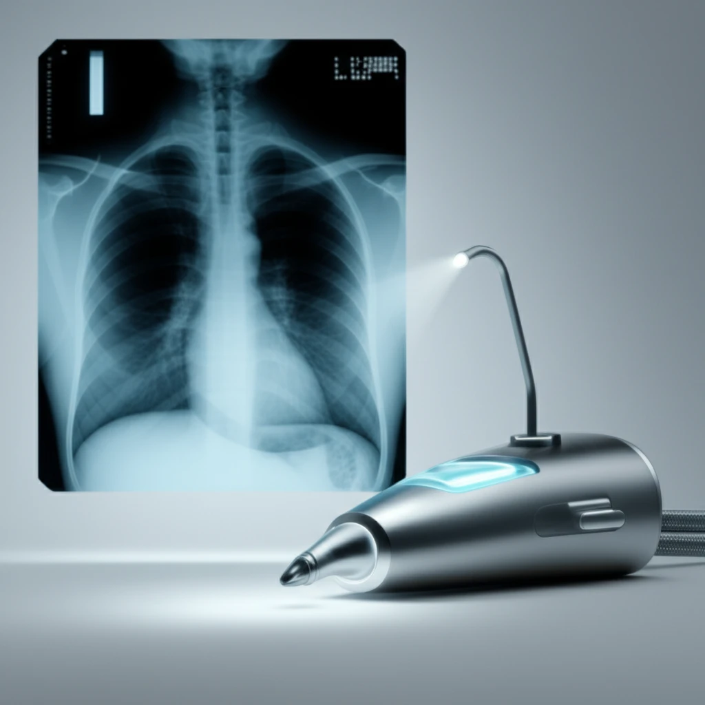
Beyond the Scalpel: Reimagining Chest X-Rays and Smoke Evacuation in Modern Medicine

"Are traditional diagnostic methods still relevant? Exploring new perspectives on chest X-rays and smoke extraction techniques."
In the fast-paced world of modern medicine, it's crucial to regularly assess the value and effectiveness of established practices. Two letters to the editor, published in the Annals of the Royal College of Surgeons of England, sparked insightful discussions regarding the utility of chest X-rays (CXRs) in diagnosing acute abdominal pain and innovative modifications to smoke evacuation devices used during surgery.
The first letter challenges a previous study questioning the necessity of routine CXRs for patients presenting with acute abdominal issues. The authors argue that CXRs remain valuable for excluding perforations and investigating respiratory symptoms, aligning with current guidelines and potentially facilitating earlier discharge for some patients. They also caution against the increased use of cross-sectional imaging, which could lead to higher costs and radiation exposure.
The second letter delves into a technical modification of a hand-held smoke extraction device, initially described by King and Ferguson. The author shares their experience with a refined technique that enhances smoke evacuation efficiency and precision during surgical procedures. This adaptation involves incising the suction tubing to allow controlled movement of the diathermy blade, reducing lumen occlusion and improving safety.
Current Role and Data Landscape
Despite the increasing use of computed tomography, chest X-ray remains the first-line investigation for suspected lung cancer in primary care. Research published in 2023 examined associations among chest X-ray trajectories, smoking, and lung-cancer risk because those relationships remained uncertain. Newer resources such as ReXGradient-160K provide large-scale datasets containing studies, images, patients, and report-text statistics, while the FDA's Nationwide Evaluation of X-Ray Trends program tracks national X-ray-use patterns.
The Standard Chest-Imaging Pathway
Chest radiography is a widely used starting point for evaluating chest disease, with interpretation focused on normal appearances and differential diagnoses when abnormalities are found. Plain chest radiography can clarify important findings, but its limitations sometimes require advanced imaging for more detailed delineation of pathology. Reviews of lung-imaging methods emphasize that each technique has particular indications, strengths, and limitations. A chest X-ray cannot show the act of smoking or nicotine itself, although it may reveal chronic structural changes associated with long-term tobacco use.
From Roentgen to Modern Radiography
Wilhelm Conrad Roentgen discovered X-rays in November 1895 after observing rays that passed through many substances and cast shadows of solid objects. The history of medical X-rays also includes earlier evidence by Pluecker and later hardware milestones such as tungsten cathodes in 1913, rotating anodes in 1929, ceramics in 1980, liquid bearings in 1990, and high-g-force technology in 2007. Radiography subsequently progressed from describing disease appearances to large clinical trials, advances in X-ray-production physics, radiation safety, and contemporary AI diagnostics.
The Ongoing Role of Chest X-Rays: Balancing Necessity and Resource Management
The debate surrounding the routine use of CXRs highlights the need for a balanced approach. While some studies suggest CXRs may not always be necessary, others emphasize their continued importance in specific clinical scenarios. The key lies in adhering to established guidelines and considering the potential consequences of omitting this diagnostic tool.
- Excluding Perforation: CXRs are effective in identifying free air in the abdomen, a sign of perforation that requires immediate surgical intervention.
- Investigating Respiratory Symptoms: In patients presenting with respiratory symptoms alongside abdominal pain, CXRs can help diagnose underlying pulmonary conditions.
- Facilitating Early Discharge: A normal CXR result may allow for earlier discharge in certain cases, reducing hospital stay and associated costs.
- Cost-Effectiveness: Compared to more advanced imaging techniques like CT scans, CXRs are a relatively inexpensive diagnostic tool.
An Evolving Evidence Base
Current research on chest X-rays and related clinical technologies is evolving, but the available evidence does not establish a single new standard for all applications. Emerging work appears to be testing how imaging, datasets, and automated interpretation might complement established practice. These developments should therefore be treated as promising areas for evaluation rather than universally validated replacements for clinical judgment.
Limits of Confidence
Chest X-rays can be useful without being definitive, and their findings may require confirmation through additional imaging or clinical assessment. Automated or innovative approaches may also perform differently across settings, patient groups, and workflow conditions. As a result, claims of greater precision or efficiency should be weighed against uncertainty, validation requirements, and the consequences of missed or ambiguous findings.
Clues Versus Confirmation
A chest X-ray cannot definitively prove that someone smokes, but it may reveal signs of lung damage or other smoking-related conditions. It is also difficult to distinguish damage from direct smoking from damage associated with secondhand-smoke exposure using a chest X-ray alone, because the resulting lung changes can be similar. The comparison highlights a central distinction: radiography may provide clinical clues, but it is not a standalone confirmation of smoking status or exposure source.
Refining Surgical Techniques: A Focus on Precision and Efficiency
The modified smoke extraction technique demonstrates the importance of continuous improvement in surgical practices. By refining existing methods, surgeons can enhance precision, reduce complications, and improve patient outcomes.
A More Careful Future
The material supports a balanced view of innovation in chest imaging and smoke evacuation. New tools may improve speed, precision, or workflow, but their value depends on how reliably they perform in real clinical environments. A responsible synthesis therefore combines technical ambition with validation, transparent limits, and continued professional oversight.
Growth in Imaging and Extraction
One market report values the global smoke-extraction-systems market at $4.8 billion in 2025 and projects it to reach $7.8 billion by 2034, representing a 6.2% compound annual growth rate. The same market is described as including natural, mechanical, and hybrid smoke-extraction systems. Separately, reported demand for efficient chest imaging, healthcare-infrastructure investment, and radiography advances is supporting global growth in digital chest-radiography systems. These projections describe market direction rather than guaranteed clinical outcomes.
Innovation Within Real Systems
The wider challenge is not simply whether a device or imaging method works in isolation, but whether it can be integrated safely into clinical workflows. Access to equipment, training, validation, maintenance, and follow-up can influence real-world performance. These systemic factors mean that technological progress alone may not produce consistent improvements across healthcare settings.
Keeping Patients and Clinicians Central
Interpreting chest X-rays is complex, and AI algorithms for this task are still being developed. External validation is important before such systems are relied upon broadly, while one retrospective single-site study compared a deep-learning algorithm with five radiologists of differing experience. Case reports and diagnostic case studies also present X-rays as tools that can contribute to breakthrough diagnoses and improved outcomes, but these examples do not remove the need for careful clinical interpretation.
The key benefits of the modified technique include:
<ul> <li><b>Improved Smoke Evacuation:</b> The modified design minimizes lumen occlusion, resulting in more effective smoke removal.</li> <li><b>Enhanced Precision:</b> Surgeons can precisely control the length of the exposed diathermy blade, allowing for targeted tissue coagulation.</li> <li><b>Increased Safety:</b> Insulating the remaining blade within the lumen reduces the risk of unintended tissue damage.</li> </ul>