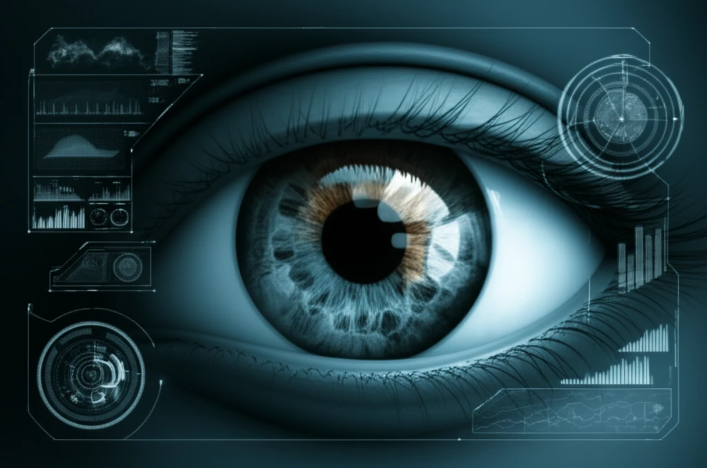
Advanced retinal scan highlighting the suprachoroidal space.

Beyond the Retina: The Emerging Potential of the Suprachoroidal Space

"Unlock new possibilities in eye health: Discover how advanced imaging and targeted drug delivery are transforming our understanding and treatment of retinal diseases through the suprachoroidal space."


The suprachoroidal space (SCS), a long-recognized potential space between the choroid and sclera, is rapidly becoming a focal point in ophthalmology. Traditionally, this space was only observable through histology or, indirectly, via ultrasonography in specific pathological conditions. However, recent advancements in optical coherence tomography (OCT) imaging are changing this landscape, offering detailed in vivo visualization.

OCT, particularly enhanced depth imaging (EDI-OCT) and swept-source OCT, has revolutionized retinal imaging, allowing clinicians to see beyond the retina and into the underlying layers. These techniques enable the identification of changes in choroidal thickness associated with various retinal diseases, including central serous chorioretinopathy, polypoidal choroidal vasculopathy, myopic degeneration, and age-related macular degeneration (AMD).

Now, with the ability to accurately image the SCS in living eyes, researchers are gaining new insights into its role in both healthy and diseased states. This enhanced visualization not only aids in diagnosis and monitoring but also opens doors for innovative medical and surgical interventions, positioning the SCS as a key area of interest for retinal specialists.

Imaging the Suprachoroidal Space: What are we seeing?

Advanced retinal scan highlighting the suprachoroidal space.

While studies on choroidal thickness often define the inner border of the sclera as the choroid's outer boundary, the presence of a hyporeflective band between the choroid and sclera has been noted. This band, now identified as the SCS, can introduce variability in choroidal thickness measurements. Advanced OCT techniques are essential for accurately visualizing and measuring this space.

Studies show the SCS can be detected in roughly half the population over 50, and its presence has been correlated with hyperopic refractive error. The exact reasons for its increased visibility with age aren't fully understood, theories suggest increased hydrostatic pressure in hyperopic eyes and protein leakage from choroidal vessels may contribute.

  • AMD: Detected in 20-50% of eyes with age-related macular degeneration.
  • Macular Holes/Epiretinal Membranes: Found in 50-60% of patients.
  • Central Serous Chorioretinopathy: Fluid loculation in the outer choroid is seen in approximately 65% of eyes.
While the significance of SCS characteristics in retinal diseases requires further investigation, monitoring SCS visibility and thickness may provide valuable diagnostic and follow-up information.

The Future of the Suprachoroidal Space: A New Frontier in Eye Care

The suprachoroidal space is garnering increased attention, and its importance in the diagnosis and treatment of various retinal conditions is becoming more evident. Recent technological advancements have enabled clear imaging of the SCS, shedding light on its variability in aging, health, and disease.

Beyond imaging, the SCS is emerging as a promising target for both drug delivery and surgical interventions. From microneedles for targeted drug administration to novel retinal prostheses, various innovative devices are being developed to leverage this space.

In the future, the SCS will likely become a routine consideration in the diagnosis, monitoring, and treatment of retinal diseases. As research continues to uncover its potential, clinicians should familiarize themselves with identifying and characterizing the SCS in OCT scans, paving the way for new insights and clinically significant advancements in eye care.

About this Article -

This article was crafted using a human-AI hybrid and collaborative approach. AI assisted our team with initial drafting, research insights, identifying key questions, and image generation. Our human editors guided topic selection, defined the angle, structured the content, ensured factual accuracy and relevance, refined the tone, and conducted thorough editing to deliver helpful, high-quality information.See our About page for more information.

This article is based on research published under:

DOI-LINK: 10.2147/opth.s89784, Alternate LINK

Title: The Suprachoroidal Space: From Potential Space To A Space With Potential

Subject: Ophthalmology

Journal: Clinical Ophthalmology

Publisher: Informa UK Limited

Authors: Elad Moisseiev, Anat Loewenstein, Glenn Yiu

Published: 2016-01-01

Everything You Need To Know

1

What exactly is the suprachoroidal space?

The suprachoroidal space (SCS) is a potential space found between the choroid and sclera in the eye. Initially, it was only observed through histology or indirectly via ultrasonography in specific pathological conditions. However, it's now being visualized directly with advanced imaging techniques such as optical coherence tomography (OCT).

2

Why is optical coherence tomography (OCT) important in understanding the suprachoroidal space?

Optical coherence tomography (OCT), especially enhanced depth imaging (EDI-OCT) and swept-source OCT, is significant because it allows detailed in vivo visualization of the suprachoroidal space (SCS). This helps in the diagnosis and monitoring of retinal diseases. By visualizing the SCS, researchers can see beyond the retina and into the underlying layers, which gives crucial diagnostic information.

3

What retinal conditions are associated with the suprachoroidal space?

The suprachoroidal space (SCS) visualization is associated with the following conditions: Age-related macular degeneration (AMD) is detected in 20-50% of the eyes. Macular Holes/Epiretinal Membranes are found in 50-60% of patients. Central Serous Chorioretinopathy has fluid loculation in the outer choroid is seen in approximately 65% of eyes. The SCS presence and characteristics can assist in diagnosis and monitoring of these diseases.

4

What causes the suprachoroidal space to be more visible with age?

The increased visibility of the suprachoroidal space (SCS) with age could be due to various factors. Research suggests that increased hydrostatic pressure in hyperopic eyes and protein leakage from choroidal vessels may contribute. The exact reasons are not fully understood. It is a topic of ongoing investigation, given that the SCS's presence is more readily detectable in a significant portion of the population over 50.

5

How might the suprachoroidal space impact the future of eye care?

The future of the suprachoroidal space (SCS) in eye care is promising. The advancements in imaging have revealed its significance in diagnosing and treating retinal conditions. Continued research into the SCS is expected to provide valuable insights into retinal diseases. It's considered a new frontier and an area of intense interest for retinal specialists because of its diagnostic and therapeutic potential.

Newsletter Subscribe

Subscribe to get the latest articles and insights directly in your inbox.