Beyond the Break: Unveiling the Secrets of Femoral Neck Fractures and Bone Health
"New research delves into the microscopic world of bone to understand why some women face hip fractures, while others don't."
Hip fractures, particularly those affecting the femoral neck, pose a significant health challenge, especially for older adults. These fractures can lead to serious complications, decreased mobility, and a reduced quality of life. While factors like osteoporosis are known contributors, the precise mechanisms behind these fractures are complex and not fully understood. Recent research, however, is shedding light on the intricate details of bone structure and how it relates to fracture risk.
This article examines a study that utilized histomorphometry, a technique that allows researchers to analyze the microscopic structure of bone tissue. The study compared the femoral necks of women over 60 who had experienced a femoral neck fracture with those who had not. By examining the bone's architecture at a cellular level, the researchers aimed to identify key differences that could explain why some individuals are more vulnerable to these debilitating fractures. The findings offer valuable insights into bone health and the factors that contribute to fracture risk.
Understanding the differences in bone structure between those who experience fractures and those who do not is crucial for developing effective preventative strategies. This research highlights the potential for early detection and intervention, emphasizing the importance of bone health monitoring and proactive measures to reduce the risk of hip fractures. The goal is to empower individuals and healthcare providers with the knowledge needed to protect bone health and promote healthy aging.
The Histomorphometry Study: Unpacking the Microscopic World of Bone
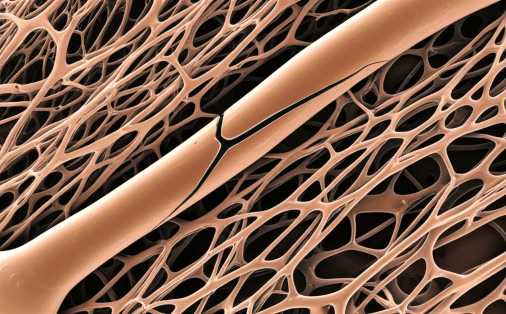
The study, published in the journal Acta Ortopédica Brasileira, employed histomorphometry to analyze the trabecular bone (spongy bone) of the femoral neck. This method involves preparing thin sections of bone tissue and examining them under a microscope to measure various structural parameters. The researchers focused on women over 60, a demographic at increased risk of hip fractures. The study aimed to identify specific characteristics of the bone that differed between those who had experienced a fracture and those who had not.
- Trabecular Thickness: The average thickness of the trabecular bone was measured, with a slight difference observed between the groups, but it was not statistically significant.
- Number of Trabeculae: The number of bone trabeculae was found to be lower in the fracture group, indicating a potential reduction in bone density.
- Trabecular Separation: The separation between the bone trabeculae was greater in the fracture group, suggesting a more porous and weaker bone structure.
- Bone Mineral Density: While bone densitometry was performed, no significant differences were observed in bone mineral density between the two groups.
Implications and Future Directions
This research provides valuable insights into the complex relationship between bone structure and femoral neck fractures. The findings highlight the importance of bone health assessments, including histomorphometry, in identifying individuals at high risk. Future research could focus on developing and refining these assessment techniques, as well as exploring targeted interventions to improve bone structure and reduce fracture risk. By understanding the microscopic details of bone, we can empower individuals to take proactive steps towards maintaining strong, healthy bones and reducing the burden of hip fractures. Further studies are needed to translate these findings into practical clinical applications, ultimately improving the lives of those at risk of these debilitating fractures.