Illustration of a kidney with a glowing aura, representing the health of a transplanted organ during a shear-wave sonoelastography procedure.

Beyond the Biopsy: How Advanced Imaging is Revolutionizing Kidney Transplant Care

"A Deep Dive into Shear-Wave Sonoelastography and Its Impact on Kidney Transplant Health"


Kidney transplantation is a life-saving procedure, offering a new beginning for individuals suffering from end-stage renal disease. However, the journey doesn't end with the transplant. The ongoing health of the transplanted kidney, or allograft, is crucial for long-term success. Regular monitoring is essential to detect any signs of dysfunction, which can lead to graft failure. Traditionally, this has involved blood tests, urine analysis, and, at times, invasive biopsies. But now, a new player has entered the field: shear-wave sonoelastography.

Shear-wave sonoelastography is a non-invasive imaging technique that provides valuable information about the stiffness of tissues. In the context of kidney transplants, this means we can now assess the health of the allograft without resorting to a biopsy in every instance. This innovative approach has the potential to significantly improve the way we monitor and manage kidney transplant patients, leading to earlier detection of problems and more effective treatment strategies.

This article will delve into the world of shear-wave sonoelastography, exploring its applications in kidney transplant care. We'll examine how it works, what it can tell us about allograft health, and the potential benefits for patients. We'll also discuss the findings of recent research and how this technology is poised to revolutionize the way we approach kidney transplant care.

Understanding Shear-Wave Sonoelastography: A Non-Invasive Window into Allograft Health

Illustration of a kidney with a glowing aura, representing the health of a transplanted organ during a shear-wave sonoelastography procedure.

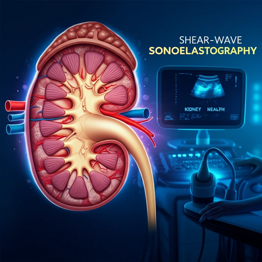
Shear-wave sonoelastography (SWSE) is a type of ultrasound imaging that measures the stiffness of tissues. It works by generating shear waves, which are mechanical waves that travel through the tissue. The speed at which these waves travel is directly related to the tissue's stiffness. Stiffer tissues, like those affected by fibrosis (scarring), will cause the waves to travel faster, while softer tissues will cause them to travel slower. This information is then used to create an image that maps the stiffness of the tissue, providing valuable insights into its health.

In the context of kidney transplants, SWSE can be used to assess the health of the allograft. By measuring the stiffness of the kidney tissue, doctors can identify signs of damage or dysfunction. This is particularly useful in detecting conditions like acute rejection, chronic allograft nephropathy (a leading cause of graft failure), and other complications. The ability to do this non-invasively is a major advantage, as it reduces the need for frequent biopsies, which are more invasive and carry a risk of complications.

  • Non-Invasive: SWSE is a completely non-invasive procedure, using ultrasound waves to assess tissue stiffness.
  • Objective Measurement: Provides objective measurements of tissue stiffness, which can be used to monitor allograft health over time.
  • Early Detection: Can help detect early signs of allograft dysfunction, allowing for timely intervention.
  • Reduced Biopsies: May reduce the need for frequent biopsies, minimizing risks and improving patient comfort.
  • Improved Monitoring: Offers a valuable tool for ongoing monitoring and management of kidney transplant patients.
Recent studies have shown promising results for SWSE in assessing kidney allograft health. These studies have demonstrated that SWSE can differentiate between stable allografts, those experiencing acute rejection, and those with chronic allograft dysfunction. The technology offers a valuable tool for clinicians to assess the functional status of the allograft, allowing them to make more informed decisions about patient care. The non-invasive nature of SWSE makes it an attractive option for routine monitoring, providing valuable information without the risks associated with more invasive procedures.

The Future of Kidney Transplant Care

Shear-wave sonoelastography represents a significant advancement in the field of kidney transplant care. Its ability to provide non-invasive assessment of allograft health opens up new possibilities for improved patient outcomes and a better quality of life. As research continues and the technology becomes more widely available, we can expect to see even greater benefits for transplant recipients. This innovative approach promises to revolutionize the way we monitor and manage kidney transplant patients, leading to a brighter future for all.

About this Article -

This article was crafted using a human-AI hybrid and collaborative approach. AI assisted our team with initial drafting, research insights, identifying key questions, and image generation. Our human editors guided topic selection, defined the angle, structured the content, ensured factual accuracy and relevance, refined the tone, and conducted thorough editing to deliver helpful, high-quality information.See our About page for more information.

This article is based on research published under:

DOI-LINK: 10.1148/radiol.2018170577, Alternate LINK

Title: Renal Allograft Dysfunction: Evaluation With Shear-Wave Sonoelastography

Subject: Radiology, Nuclear Medicine and imaging

Journal: Radiology

Publisher: Radiological Society of North America (RSNA)

Authors: Nitin P. Ghonge, Mohita Mohan, Vikas Kashyap, Sanjiv Jasuja

Published: 2018-07-01

Everything You Need To Know

1

What is shear-wave sonoelastography, and how does it aid in monitoring kidney transplant health?

Shear-wave sonoelastography (SWSE) is an ultrasound imaging technique that assesses tissue stiffness. By measuring how shear waves travel through the kidney allograft, SWSE can identify signs of damage or dysfunction, such as those related to acute rejection or chronic allograft nephropathy. Stiffer tissues cause waves to travel faster, while softer tissues cause the waves to travel slower. This information allows doctors to monitor the health of the transplanted kidney without always resorting to invasive biopsies.

2

How does shear-wave sonoelastography compare to traditional methods like biopsies in monitoring kidney transplant health?

Traditional methods for monitoring kidney transplant health often involve blood tests, urine analysis, and biopsies. Shear-wave sonoelastography (SWSE) offers a non-invasive alternative to frequent biopsies. While biopsies are invasive and carry risks, SWSE uses ultrasound waves to assess the stiffness of the allograft, providing valuable information about its health without the need for tissue extraction. This can reduce the need for biopsies, improving patient comfort and minimizing risks.

3

What are the key advantages of using shear-wave sonoelastography for kidney transplant monitoring?

The key advantages of shear-wave sonoelastography (SWSE) include its non-invasive nature, which eliminates the risks associated with biopsies, such as bleeding and infection. SWSE provides objective measurements of tissue stiffness, enabling consistent monitoring of allograft health over time. It also facilitates the early detection of allograft dysfunction, allowing for timely interventions and improved patient outcomes. Ultimately, SWSE serves as a valuable tool for ongoing monitoring and management of kidney transplant patients.

4

How can shear-wave sonoelastography differentiate between various conditions affecting a kidney allograft?

Shear-wave sonoelastography (SWSE) can differentiate between various conditions affecting a kidney allograft by measuring tissue stiffness. Studies have shown that SWSE can distinguish between stable allografts, those experiencing acute rejection, and those with chronic allograft nephropathy. This differentiation is possible because each condition alters the stiffness of the kidney tissue in specific ways. By identifying these differences, clinicians can more accurately assess the functional status of the allograft and make informed treatment decisions.

5

What impact could shear-wave sonoelastography have on the future of kidney transplant care?

Shear-wave sonoelastography (SWSE) has the potential to revolutionize kidney transplant care by providing a non-invasive method for assessing allograft health. As research continues and the technology becomes more accessible, SWSE promises to improve patient outcomes and quality of life. Its ability to detect early signs of dysfunction and reduce the need for invasive biopsies could lead to more effective treatment strategies and a brighter future for kidney transplant recipients. Furthermore, advancements in SWSE technology may lead to even more precise and comprehensive assessments of allograft health, potentially incorporating other imaging modalities for a holistic approach.

Newsletter Subscribe

Subscribe to get the latest articles and insights directly in your inbox.