A 3D-printed model of the pterygopalatine fossa, highlighting its complex structure and educational value.

Beyond Textbooks: How 3D Printing Is Revolutionizing Anatomy Education

"From Complex Structures to Hands-On Learning: Discover How 3D Printing Models Are Transforming the Way We Study Anatomy."


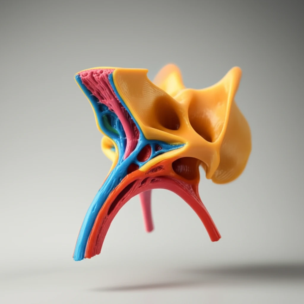
Anatomy, the cornerstone of medical understanding, has traditionally relied on textbooks, illustrations, and cadaveric dissections. These methods, while essential, often present challenges in visualizing and understanding complex, three-dimensional structures. The intricate details of areas like the pterygopalatine fossa, a critical region in the skull, are difficult to grasp using conventional methods. However, a technological revolution is underway, transforming the way we learn and teach anatomy.

3D printing, also known as additive manufacturing, is emerging as a powerful tool in medical education. This technology allows for the creation of tangible models from digital data, offering a more accessible and immersive learning experience. Imagine being able to hold a perfect replica of the pterygopalatine fossa in your hands, exploring its intricate canals and relationships in a way that textbooks and diagrams simply can't replicate.

This article explores the innovative application of 3D printing in anatomy education, specifically focusing on the creation of a 3D-printed model of the pterygopalatine fossa. We'll delve into the process, benefits, and potential of this groundbreaking approach, highlighting how it's changing the landscape of anatomical learning for students and professionals alike.

Unveiling the Pterygopalatine Fossa: The Challenge of Traditional Anatomy Study

A 3D-printed model of the pterygopalatine fossa, highlighting its complex structure and educational value.

The pterygopalatine fossa (PPF) is a small but crucial space located in the skull base. It's a complex region housing important nerves and blood vessels. Understanding the PPF is essential for medical professionals, including surgeons, radiologists, and dentists. Its intricate structure, with its canals, foramina, and surrounding structures, has always been a challenge to grasp.

Traditional learning methods often fall short in conveying the true complexity of this region. Cadaveric dissections, while offering hands-on experience, can be limited by access, preservation challenges, and the two-dimensional nature of textbooks and illustrations. Students often struggle to mentally reconstruct the three-dimensional relationships within the PPF using these tools alone.

  • Limited Visualization: Textbooks and diagrams offer a simplified 2D representation.
  • Accessibility: Cadaveric dissections are not always readily available.
  • Complexity: The intricate structure of the PPF is challenging to understand.
The introduction of 3D printing offers a powerful solution to these limitations, providing a new way to explore and understand the PPF and other complex anatomical structures.

The Future of Anatomy Education: 3D Printing and Beyond

3D printing is poised to play an even more significant role in anatomy education. As technology advances and costs decrease, the use of 3D-printed models will likely become even more widespread. The ability to create personalized models, incorporate haptic feedback, and integrate with augmented reality will further enhance the learning experience. By embracing these innovative tools, we can create a new generation of medical professionals with a deeper understanding of the human body and the ability to provide better patient care.

About this Article -

This article was crafted using a human-AI hybrid and collaborative approach. AI assisted our team with initial drafting, research insights, identifying key questions, and image generation. Our human editors guided topic selection, defined the angle, structured the content, ensured factual accuracy and relevance, refined the tone, and conducted thorough editing to deliver helpful, high-quality information.See our About page for more information.

This article is based on research published under:

DOI-LINK: 10.1007/s00276-017-1916-x, Alternate LINK

Title: 3D Printing The Pterygopalatine Fossa: A Negative Space Model Of A Complex Structure

Subject: Radiology, Nuclear Medicine and imaging

Journal: Surgical and Radiologic Anatomy

Publisher: Springer Science and Business Media LLC

Authors: Ross Bannon, Shivani Parihar, Yiannis Skarparis, Ourania Varsou, Enis Cezayirli

Published: 2017-08-30

Everything You Need To Know

1

How is 3D printing revolutionizing anatomy education?

3D printing is revolutionizing anatomy education by providing detailed, hands-on models of complex structures. Instead of relying solely on textbooks and illustrations, students can now interact with tangible, three-dimensional representations of anatomical features, like the pterygopalatine fossa, enhancing understanding and retention. This technology allows for a more immersive and accessible learning experience, making it easier to grasp intricate details and spatial relationships.

2

What are the limitations of traditional methods like textbooks and cadaveric dissections in studying anatomy?

Traditional methods, such as textbooks and cadaveric dissections, present several limitations. Textbooks and diagrams offer simplified, two-dimensional representations, which can make it difficult to visualize complex, three-dimensional structures. Cadaveric dissections, while offering hands-on experience, are limited by accessibility, preservation challenges, and the inherent difficulties in mentally reconstructing anatomical relationships. For instance, grasping the intricacies of the pterygopalatine fossa is particularly challenging with these methods.

3

What is the pterygopalatine fossa, and why is it important for medical professionals?

The pterygopalatine fossa (PPF) is a small but crucial space located in the skull base, housing important nerves and blood vessels. Understanding the PPF is essential for medical professionals, including surgeons, radiologists, and dentists, due to its involvement in various cranial procedures and the potential for pathologies that affect the area. Its intricate structure, with canals, foramina, and surrounding structures, makes it a challenging region to understand, highlighting the need for innovative learning tools.

4

How does 3D printing improve the study of the pterygopalatine fossa specifically?

3D printing significantly improves the study of the pterygopalatine fossa (PPF) by allowing the creation of detailed, tangible models. Students can physically interact with these models, exploring the intricate canals, foramina, and surrounding structures in a way that textbooks and diagrams simply can't replicate. This hands-on approach enhances understanding and retention, providing a more immersive and accessible learning experience compared to traditional methods.

5

What is the future of 3D printing in anatomy education, and what advancements are expected?

The future of 3D printing in anatomy education is promising, with advancements expected to further enhance the learning experience. As technology advances and costs decrease, 3D-printed models will likely become even more widespread. Future developments include the ability to create personalized models, incorporate haptic feedback for a more realistic touch, and integrate with augmented reality to overlay digital information onto physical models. These innovations will enable a deeper understanding of the human body and better patient care.

Newsletter Subscribe

Subscribe to get the latest articles and insights directly in your inbox.