Microscopic view of Auer rods within a blood vessel.

Auer Rods: What They Indicate About Your Health

"Unveiling the Secrets Hidden in Blood Smears"


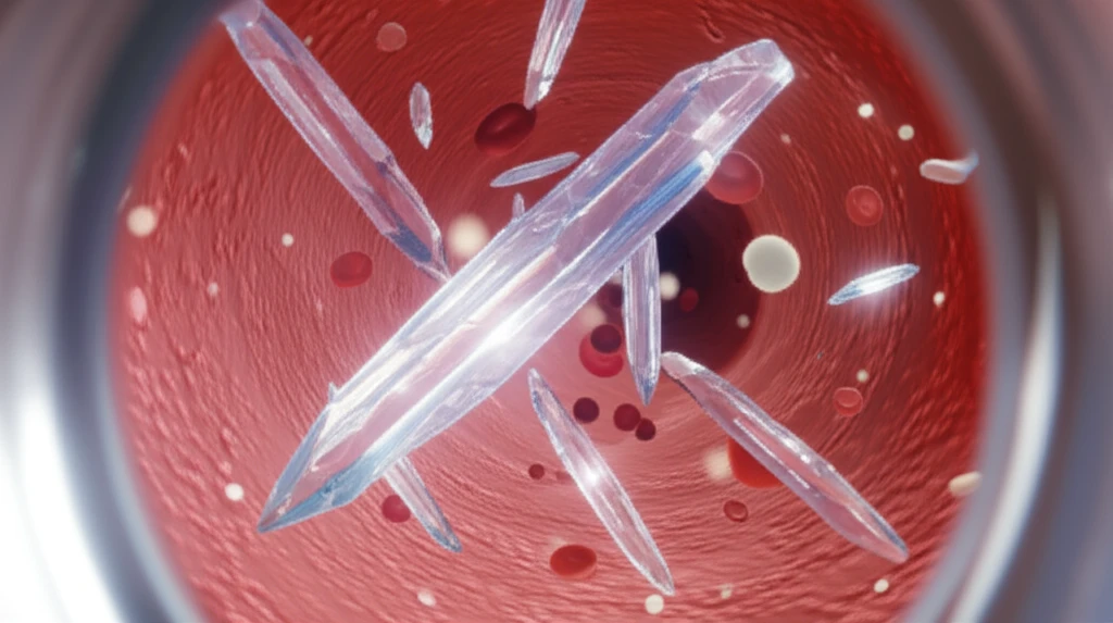
Imagine peering through a microscope, not at some distant galaxy, but at the very essence of life: blood. Within this intricate landscape, pathologists and hematologists search for clues, tiny anomalies that can unlock the secrets of a patient's health. Among these microscopic findings are Auer rods, crystalline structures found within the cytoplasm of certain white blood cells. Their presence, though not always a cause for alarm, often signals a deeper, more complex medical condition that warrants investigation.

Auer rods aren't a normal component of blood cells. They are abnormal inclusions, essentially crystal-like formations of fused lysosomes (cellular digestive units) that appear as slender, rod-shaped structures within the cytoplasm of myeloid blast cells. These cells are precursors to mature white blood cells, specifically granulocytes like neutrophils, eosinophils, and basophils. The discovery of Auer rods, named after the American physiologist and hematologist John Auer, is a crucial diagnostic indicator in certain types of blood cancers, particularly acute myeloid leukemia (AML).

This article will delve into the world of Auer rods, exploring their significance in diagnosing and understanding various hematological conditions. We'll unpack the science behind their formation, the diseases they're associated with, and what their presence means for patients and their healthcare journey. Whether you're a medical professional seeking a refresher or simply curious about the microscopic world within us, this guide offers a comprehensive overview of Auer rods and their clinical implications.

What are Auer Rods and How Do They Form?

Microscopic view of Auer rods within a blood vessel.

Auer rods are essentially abnormal clumps of azurophilic granules – specialized lysosomes that contain enzymes and proteins. These granules normally play a role in the immune response, helping white blood cells destroy bacteria and other pathogens. In certain conditions, however, these granules fuse together, forming larger, elongated crystals that are visible under a microscope. These crystals are what we know as Auer rods.

The formation of Auer rods is primarily associated with disruptions in the normal maturation process of myeloid cells. In healthy individuals, myeloid cells develop in a carefully orchestrated sequence, differentiating into mature granulocytes. However, in conditions like AML, this process is disrupted, leading to the accumulation of immature blast cells in the bone marrow and blood. These blast cells often contain Auer rods due to the abnormal fusion of their azurophilic granules.

  • Disrupted Maturation: In conditions such as AML, the normal maturation of myeloid cells is interrupted, leading to an accumulation of immature blast cells.
  • Abnormal Fusion: Within these blast cells, azurophilic granules (specialized lysosomes) abnormally fuse together.
  • Crystal Formation: This fusion results in the formation of elongated, crystal-like structures known as Auer rods.
  • Microscopic Visibility: These rods are visible under a microscope, aiding in the diagnosis of certain blood disorders.
It's important to note that Auer rods are not found in all types of leukemia. They are primarily associated with AML, particularly certain subtypes like acute promyelocytic leukemia (APL). Their presence can help distinguish AML from other types of leukemia and guide treatment decisions. Moreover, their absence doesn't automatically rule out AML, as some AML subtypes may not exhibit Auer rods.

The Future of Auer Rod Research

While Auer rods have been recognized as a diagnostic marker for decades, research continues to refine our understanding of their formation, composition, and clinical significance. Ongoing studies are exploring the specific molecular mechanisms that lead to granule fusion, as well as the potential for targeting these pathways for therapeutic intervention. Furthermore, advancements in microscopy and imaging techniques are enabling more detailed visualization and analysis of Auer rods, potentially leading to improved diagnostic accuracy and personalized treatment strategies. The microscopic world of Auer rods continues to offer valuable insights into the complexities of blood disorders and the ongoing quest for better treatments.

About this Article -

This article was crafted using a human-AI hybrid and collaborative approach. AI assisted our team with initial drafting, research insights, identifying key questions, and image generation. Our human editors guided topic selection, defined the angle, structured the content, ensured factual accuracy and relevance, refined the tone, and conducted thorough editing to deliver helpful, high-quality information.See our About page for more information.

Everything You Need To Know

1

What exactly are Auer rods, and what do they look like under a microscope?

Auer rods are abnormal, crystalline structures found within the cytoplasm of myeloid blast cells. They appear as slender, rod-shaped structures when viewed under a microscope. These formations are essentially fused azurophilic granules, which are specialized lysosomes containing enzymes and proteins. These granules clump together abnormally, forming the distinctive crystal-like Auer rods, a key diagnostic indicator in certain blood disorders.

2

In what specific blood disorders are Auer rods most commonly observed, and why is this important?

Auer rods are primarily associated with acute myeloid leukemia (AML), particularly subtypes like acute promyelocytic leukemia (APL). Their presence is crucial because it helps differentiate AML from other types of leukemia, guiding treatment decisions. The identification of Auer rods in a blood smear provides essential information for hematologists and oncologists to accurately diagnose the specific type of leukemia and tailor the most effective treatment strategy.

3

How do Auer rods form within cells, and what does this tell us about the underlying cellular processes?

Auer rods form due to a disruption in the normal maturation process of myeloid cells. In conditions like AML, this process is interrupted, leading to the accumulation of immature blast cells. Within these blast cells, azurophilic granules, which are normally involved in the immune response, abnormally fuse together. This fusion results in the formation of the elongated, crystal-like structures known as Auer rods. This indicates a failure in the normal differentiation and maturation pathways of myeloid cells, a hallmark of AML.

4

What is the significance of the presence or absence of Auer rods in diagnosing blood disorders, and what are the limitations?

The presence of Auer rods is a strong indicator of AML, aiding in its diagnosis. However, their absence does not necessarily rule out AML, as some subtypes may not exhibit these rods. Additionally, Auer rods are not found in all types of leukemia. Therefore, while their presence is diagnostically significant, their absence requires further investigation and other diagnostic tests to determine the underlying cause of the blood disorder. It's essential to consider other clinical findings and laboratory results for a complete diagnosis.

5

What are the future directions in Auer rod research, and how might this improve the diagnosis and treatment of blood disorders?

Future research on Auer rods focuses on understanding the molecular mechanisms of granule fusion and exploring potential therapeutic targets. Scientists are studying how the azurophilic granules fuse and whether there are specific pathways that can be targeted to prevent their formation. Advancements in microscopy and imaging techniques also allow for more detailed analysis of Auer rods. These advances could improve diagnostic accuracy, leading to more personalized treatment strategies for patients with AML and other related blood disorders, ultimately improving outcomes.

Newsletter Subscribe

Subscribe to get the latest articles and insights directly in your inbox.