Illustration depicting Chilaiditi syndrome, showing the colon between the liver and diaphragm on an X-ray.

Air Under the Diaphragm? When It's NOT an Emergency

"Discover the rare Chilaiditi syndrome, a condition that mimics a surgical emergency but often requires only supportive care."


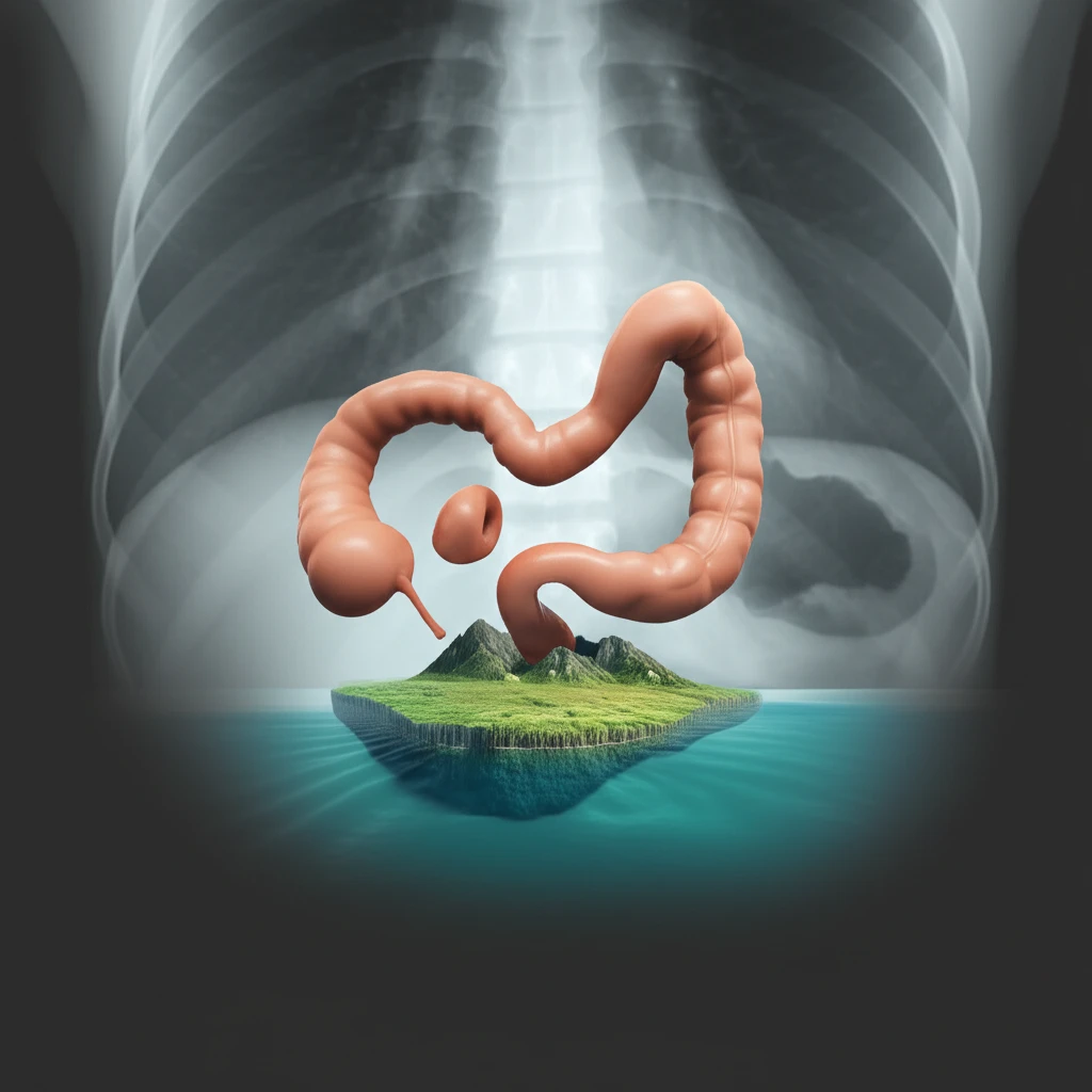
When doctors spot air under the diaphragm in an X-ray, it's usually a red alert, suggesting a potentially life-threatening situation that needs immediate surgery. However, there are exceptions. This article will shed light on a rare condition known as Chilaiditi syndrome, a diagnostic surprise that every healthcare professional should be aware of.

Chilaiditi sign, the hallmark of this syndrome, is a radiologic finding where a portion of the colon slips between the liver and the diaphragm. In most instances, it’s discovered incidentally during imaging for other reasons. Yet, some patients experience symptoms, transforming the sign into Chilaiditi syndrome. This article aims to increase familiarity with Chilaiditi syndrome, emphasize the importance of a thorough physical examination, and appropriate use of radiological tests when evaluating patients.

This article uses a case report of a 49-year-old Egyptian man who presented to the emergency department with cough and abdominal discomfort. His case, diagnosed as Chilaiditi syndrome, highlights the importance of considering this rare condition to avoid unnecessary invasive procedures.

The Case: Cough, Discomfort, and a Surprising X-Ray

Illustration depicting Chilaiditi syndrome, showing the colon between the liver and diaphragm on an X-ray.

A 49-year-old Egyptian man went to the emergency department with a 48-hour history of coughing, producing a small amount of sputum, which was causing abdominal discomfort. While he reported fatigue, he denied other symptoms such as chest pain, fever, or gastrointestinal issues. He had a history of obesity and had undergone a laparoscopic Roux-en-Y gastric bypass for weight loss, but otherwise, his medical history was unremarkable.

Upon examination, his vitals were stable, though his blood pressure was slightly elevated. While he reported mild distress, his respiratory and cardiovascular systems appeared normal. His abdomen was soft and non-tender. Initial lab results were unremarkable, but a chest X-ray revealed air under the right diaphragm – a finding that usually prompts immediate concern.

  • The Twist: Further investigation with a CT scan revealed that the 'air' was actually a loop of colon positioned between the liver and diaphragm, confirming Chilaiditi sign.
  • The Diagnosis: Based on the radiologic findings and the patient’s symptoms, a diagnosis of Chilaiditi syndrome was made.
  • The Treatment: Instead of surgery, the patient was treated with IV fluids, cough suppressants, and pain management. His pain subsided, and he was discharged in stable condition. A year later, he remained asymptomatic.
This case demonstrates the importance of considering Chilaiditi syndrome when encountering air under the diaphragm. While pneumoperitoneum typically signals a surgical emergency, Chilaiditi syndrome can be managed conservatively, avoiding unnecessary interventions.

Treat the Patient, Not Just the Image

Medical training emphasizes that air under the diaphragm often necessitates immediate surgical intervention. While this remains a crucial principle, this case underscores the need to correlate radiological findings with a comprehensive clinical assessment.

A thorough physical examination, devoid of peritonitis signs, warrants further investigation to understand the underlying pathology. By considering conditions like Chilaiditi syndrome, physicians can avoid exposing patients to unnecessary surgeries and their associated risks.

Physicians should broaden their awareness regarding potential causes of pneumoperitoneum that might not require surgical intervention. This can help prevent unnecessary surgeries, thus reducing patient risk and optimizing healthcare resource allocation.

About this Article -

This article was crafted using a human-AI hybrid and collaborative approach. AI assisted our team with initial drafting, research insights, identifying key questions, and image generation. Our human editors guided topic selection, defined the angle, structured the content, ensured factual accuracy and relevance, refined the tone, and conducted thorough editing to deliver helpful, high-quality information.See our About page for more information.

This article is based on research published under:

DOI-LINK: 10.1186/s13256-018-1804-y, Alternate LINK

Title: Chilaiditi Syndrome – A Rare Case Of Pneumoperitoneum In The Emergency Department: A Case Report

Subject: General Medicine

Journal: Journal of Medical Case Reports

Publisher: Springer Science and Business Media LLC

Authors: Mohamed M. Gad, Muneer J. Al-Husseini, Sami Salahia, Anas M. Saad, Ramy Amin

Published: 2018-09-16

Everything You Need To Know

1

What exactly is Chilaiditi syndrome?

Chilaiditi syndrome is a rare condition where a portion of the colon is positioned between the liver and the diaphragm. This is often discovered incidentally during imaging for other reasons. In this context, it's important because it can mimic a surgical emergency due to the presence of air under the diaphragm on an X-ray. However, unlike a typical pneumoperitoneum (air in the abdominal cavity), Chilaiditi syndrome often doesn't require surgery, but rather supportive care, making accurate diagnosis crucial.

2

What is the Chilaiditi sign, and why is it important?

The hallmark of Chilaiditi syndrome is the Chilaiditi sign, a radiologic finding where a portion of the colon is found between the liver and the diaphragm. It's significant because it can be mistaken for free air in the abdomen (pneumoperitoneum), which usually indicates a serious condition requiring immediate surgery. This misdiagnosis can lead to unnecessary invasive procedures. Recognizing the Chilaiditi sign allows for a more appropriate and conservative treatment approach.

3

Why is air under the diaphragm a concern, and how does Chilaiditi syndrome fit into that?

A chest X-ray revealing air under the diaphragm usually prompts immediate concern, as it often indicates pneumoperitoneum, potentially signaling a life-threatening situation that requires immediate surgery. However, in cases of Chilaiditi syndrome, the ‘air’ is actually a loop of colon positioned between the liver and diaphragm. Therefore, while air under the diaphragm is a critical finding, it's essential to correlate radiological findings with a thorough clinical assessment to differentiate between pneumoperitoneum and Chilaiditi syndrome.

4

How was the case of the 49-year-old Egyptian man relevant in this context?

In the case of the 49-year-old Egyptian man, the diagnosis of Chilaiditi syndrome was made based on the radiologic findings (the Chilaiditi sign) and the patient's symptoms. Because of this diagnosis, instead of surgery, the patient was treated with IV fluids, cough suppressants, and pain management, and was discharged in stable condition. This demonstrates that, in the context, the correct diagnosis of Chilaiditi syndrome can avoid unnecessary interventions.

5

What are the implications of being diagnosed with Chilaiditi syndrome?

If you are experiencing symptoms of Chilaiditi syndrome, the first step is a thorough physical examination. Then, appropriate radiological tests, such as a CT scan, should be used to correlate findings with a comprehensive clinical assessment. In contrast to pneumoperitoneum, which typically requires surgery, Chilaiditi syndrome can often be managed conservatively with supportive care, such as IV fluids, cough suppressants, and pain management. Therefore, if diagnosed the implications are a possibly less invasive approach to treatment and management.

Newsletter Subscribe

Subscribe to get the latest articles and insights directly in your inbox.