Surreal knee joint illustration with ACL highlighted in MRI imagery.

ACL Injury Imaging: Spotting the Pitfalls to Ensure Accurate Diagnosis

"Navigating the complexities of MRI for acute anterior cruciate ligament (ACL) injuries to improve diagnostic accuracy and treatment planning."


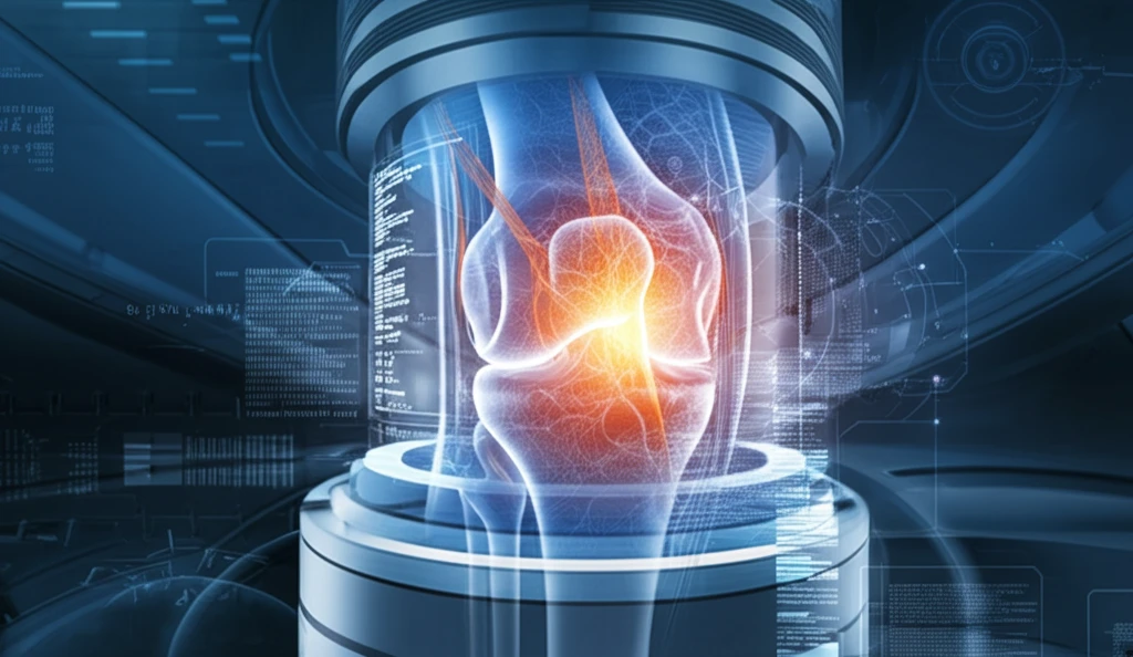
Accurate diagnosis of anterior cruciate ligament (ACL) injuries is crucial for effective treatment and rehabilitation. Magnetic resonance imaging (MRI) has become an indispensable tool for assessing these injuries. The goal of MRI is to give surgeons the most precise information on where a tear is located and the quality of the remaining tissue. This information is key to deciding whether an ACL repair is feasible, especially when considering minimally invasive arthroscopic techniques.

However, correctly reading MR images of ACL injuries isn't always straightforward. A letter to the editor regarding the publication, “Preoperative magnetic resonance imaging predicts eligibility for arthroscopic primary anterior cruciate ligament repair,” highlights the importance of carefully considering potential pitfalls that can lead to misdiagnosis. These pitfalls primarily concern the precise determination of tear location and the evaluation of tissue quality.

This article explores the challenges associated with MR imaging of acute ACL injuries, emphasizing the need for clinicians and radiologists to stay vigilant to ensure that patients receive the best possible care based on reliable imaging assessments. By understanding these challenges, healthcare providers can improve diagnostic accuracy, leading to more effective treatment plans.

Decoding ACL Tears: Why MRI Interpretation Isn't Always Clear-Cut

Surreal knee joint illustration with ACL highlighted in MRI imagery.

While MRI is generally reliable, it has limitations when dealing with ACL injuries. It is often assumed that MRI can clearly show the location of an ACL tear, guiding treatment plans. However, research indicates that in many cases, MRI results can be ambiguous. Studies show that a clear gap indicating a complete ACL tear is only visible in about 75% of cases. This means that relying solely on this sign can lead to missed diagnoses or incorrect assessments of the injury's severity.

A common challenge in diagnosing ACL tears with MRI is the presence of edema and hemorrhage, which can obscure the actual tear site. Instead of a clear tear, the MRI might show a cloudy or swollen area where the ACL should be. This is because after an ACL injury, bleeding and fluid accumulation can distort the usual appearance of the ligament. This type of presentation is quite common, with some studies reporting it in up to 88% of cases. The presence of these obscuring factors makes it hard to accurately locate and grade the tear, impacting decisions about treatment strategies.

Here are some critical points regarding the challenges in MRI interpretation of ACL injuries:
  • Discontinuity Visibility: Clear gaps indicating a complete tear are not always evident on MRI.
  • Signal and Morphology: Abnormal signals and changes in the ACL's shape due to edema and hemorrhage can obscure the exact tear location.
  • Subjectivity in Grading: Assessing tissue quality as good, fair, or poor is subjective and can vary among radiologists.
To illustrate the difficulties in accurately assessing ACL tears via MRI, consider a case where a patient’s initial MRI suggested a complete ACL tear. The images showed the typical signs of diffuse hyperintensity and non-visualization of fibers. However, during arthroscopic examination, the tear was found to be only partial. A follow-up MRI five months later showed that the ACL had regained much of its normal continuity and signal intensity as the initial edema and hemorrhage subsided. This example underscores the potential for misinterpretation in the acute setting.

Enhancing Diagnostic Precision for Better Patient Outcomes

In conclusion, while MRI is indispensable for evaluating ACL injuries, clinicians must be aware of its limitations, particularly in the acute setting. Edema and hemorrhage can obscure tear location, leading to potential misdiagnoses. Being aware of these pitfalls allows for a more nuanced interpretation of MRI results, improving diagnostic accuracy and ensuring that patients receive the most appropriate and effective treatment strategies tailored to their specific needs.

About this Article -

This article was crafted using a human-AI hybrid and collaborative approach. AI assisted our team with initial drafting, research insights, identifying key questions, and image generation. Our human editors guided topic selection, defined the angle, structured the content, ensured factual accuracy and relevance, refined the tone, and conducted thorough editing to deliver helpful, high-quality information.See our About page for more information.

This article is based on research published under:

DOI-LINK: 10.1007/s00167-017-4701-9, Alternate LINK

Title: Pitfalls In Mr Imaging Of Acute Anterior Cruciate Ligament Injuries

Subject: Orthopedics and Sports Medicine

Journal: Knee Surgery, Sports Traumatology, Arthroscopy

Publisher: Springer Science and Business Media LLC

Authors: Pieter Van Dyck

Published: 2017-09-04

Everything You Need To Know

1

Why is accurate diagnosis of ACL injuries so crucial?

Accurate diagnosis of Anterior Cruciate Ligament (ACL) injuries is essential for effective treatment and rehabilitation. It directly impacts the choice of treatment, determining whether an ACL repair is feasible, especially when considering minimally invasive arthroscopic techniques. Precise information on the tear's location and the quality of the remaining tissue guides surgeons in making informed decisions, ultimately leading to better patient outcomes.

2

What are the primary challenges in interpreting MRI results for ACL injuries?

The main challenges in interpreting Magnetic Resonance Imaging (MRI) results for Anterior Cruciate Ligament (ACL) injuries include the ambiguity in identifying complete tears and the presence of edema and hemorrhage. Clear gaps indicating complete tears are not always visible; they are apparent in approximately 75% of cases. Edema and hemorrhage can obscure the exact tear location, potentially leading to misdiagnosis of the injury's severity or location. Subjectivity in grading tissue quality (good, fair, or poor) adds further complexity and potential variability in diagnosis among radiologists.

3

How can edema and hemorrhage affect the accuracy of an MRI in diagnosing an ACL injury?

Edema and hemorrhage can significantly impact the accuracy of MRI in diagnosing Anterior Cruciate Ligament (ACL) injuries by obscuring the actual tear site. Instead of a clear indication of a tear, the MRI might show a cloudy or swollen area where the ACL should be. This is because bleeding and fluid accumulation can distort the typical appearance of the ligament. This obscures the ability to accurately locate and grade the tear, potentially affecting decisions about the treatment strategy. Some studies have shown the presence of these obscuring factors in up to 88% of cases.

4

What are the implications of relying solely on MRI results for ACL tear diagnosis?

Relying solely on Magnetic Resonance Imaging (MRI) results for Anterior Cruciate Ligament (ACL) tear diagnosis can lead to missed diagnoses or incorrect assessments of the injury's severity. For instance, the absence of a clear gap indicative of a complete tear in 25% of cases may lead to an incomplete diagnosis. Considering the presence of edema and hemorrhage, which can obscure the tear location, further complicates accurate assessment. Such inaccuracies can lead to inappropriate treatment plans, which emphasizes the need for a more nuanced interpretation of MRI results, considering all potential pitfalls.

5

What steps can be taken to improve diagnostic accuracy when evaluating ACL injuries using MRI?

To improve diagnostic accuracy when evaluating Anterior Cruciate Ligament (ACL) injuries using Magnetic Resonance Imaging (MRI), clinicians and radiologists must be aware of the limitations of MRI, especially in the acute setting. This involves carefully considering potential pitfalls, particularly the impact of edema and hemorrhage on obscuring tear location. A more nuanced interpretation of MRI results is essential, which means not relying solely on visible gaps. This enables healthcare providers to improve diagnostic accuracy and develop more effective treatment plans tailored to specific patient needs, which leads to better patient outcomes.

Newsletter Subscribe

Subscribe to get the latest articles and insights directly in your inbox.