Illustration of a torn ACL within a knee joint, overlaid with MRI scan lines.

ACL Injury Imaging: Are You Seeing the Whole Picture?

"Uncover the hidden pitfalls in MRI diagnosis of acute ACL tears and learn how to avoid misinterpretations."


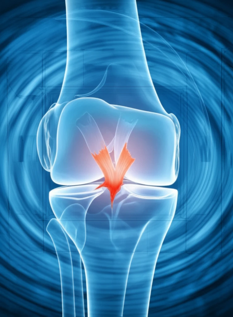
Anterior cruciate ligament (ACL) injuries are a common problem, especially among athletes. When someone suspects an ACL tear, magnetic resonance imaging (MRI) is often the first line of defense to confirm the diagnosis. MRI helps doctors visualize the soft tissues inside the knee, including the ACL. This imaging guides decisions about whether surgery is needed and how best to approach the repair.

However, MRI isn't always a slam dunk. Interpreting these images can be tricky, and sometimes, the initial MRI might not give the full story. This can lead to uncertainty about the extent of the injury and potentially affect treatment plans.

This article dives into the potential pitfalls of using MRI to assess acute ACL injuries. We'll explore some common misinterpretations and highlight the factors that can make accurate diagnosis challenging. Ultimately, the goal is to help you understand the limitations of MRI and ensure you're equipped to make informed decisions about your knee health.

The MRI Illusion: Why ACL Tears Aren't Always Clear-Cut

Illustration of a torn ACL within a knee joint, overlaid with MRI scan lines.

When an ACL tears, it doesn't always result in a clean, obvious break that's easily spotted on an MRI. In many cases, the injury presents with subtle signs like swelling (edema), bleeding (hemorrhage), and a general disruption of the ligament's normal appearance. These factors can obscure the actual tear, making it difficult to determine the precise location and severity of the damage.

One study found that a clear gap in the ACL, typically considered a primary sign of a complete tear, is only visible in about 75% of cases. This means that in a significant number of injuries, the MRI might not show the classic signs you'd expect. Instead, the ACL might appear as a blurry, undefined mass due to the surrounding edema and hemorrhage.

  • Swelling and Bleeding: Post-injury inflammation can cloud the image.
  • Partial Tears: Distinguishing between partial and complete tears can be challenging.
  • Individual Variation: Everyone's anatomy is slightly different, making it harder to define "normal."
Consider a real-life scenario: An MRI scan taken shortly after an ACL injury shows a disrupted ligament with significant swelling. It's difficult to pinpoint the exact location or extent of the tear. During arthroscopic surgery, performed to address a meniscal issue, the surgeon discovers a partial ACL tear. A follow-up MRI months later, after the swelling has subsided, reveals that the ACL has regained some continuity and a more normal appearance. This case illustrates how the initial post-trauma conditions can mask the true extent of the injury on MRI.

MRI: A Powerful Tool, But Not the Only One

MRI remains an invaluable tool for evaluating ACL injuries. However, it's important to recognize its limitations and avoid relying solely on MRI findings. Clinical examination, including physical tests performed by an experienced orthopedic surgeon or sports medicine physician, plays a crucial role in assessing the stability of the knee and the extent of the injury. In some cases, further imaging or even diagnostic arthroscopy may be necessary to get a complete picture.

About this Article -

This article was crafted using a human-AI hybrid and collaborative approach. AI assisted our team with initial drafting, research insights, identifying key questions, and image generation. Our human editors guided topic selection, defined the angle, structured the content, ensured factual accuracy and relevance, refined the tone, and conducted thorough editing to deliver helpful, high-quality information.See our About page for more information.

Everything You Need To Know

1

What is the primary role of MRI in diagnosing Anterior Cruciate Ligament (ACL) injuries?

MRI is often the first imaging method used to visualize the soft tissues inside the knee, particularly the Anterior Cruciate Ligament (ACL). Its primary role is to confirm the diagnosis of an ACL tear, providing crucial information to guide decisions on whether surgery is needed and to plan the approach for the repair. By showing the condition of the ACL and surrounding structures, MRI aids in forming an effective treatment strategy.

2

Why can't an MRI always clearly identify an Anterior Cruciate Ligament (ACL) tear?

An MRI does not always provide a clear view of an Anterior Cruciate Ligament (ACL) tear due to several factors. Swelling (edema) and bleeding (hemorrhage) that occur after an injury can obscure the tear, making it difficult to pinpoint the exact location and severity of the damage. The ACL might appear as a blurry mass instead of a clear break. The presence of partial tears, and individual variations in anatomy further complicate the interpretation of MRI images, potentially leading to misinterpretations regarding the extent of the injury.

3

What are some common challenges in interpreting MRI scans for Anterior Cruciate Ligament (ACL) injuries?

Several challenges can arise when interpreting MRI scans for Anterior Cruciate Ligament (ACL) injuries. One key challenge is the presence of swelling (edema) and bleeding (hemorrhage), which can cloud the image and obscure the tear's appearance. Distinguishing between partial and complete tears can also be difficult, requiring careful analysis. Additionally, individual anatomical variations mean that what is 'normal' can differ from person to person, making it harder to define the injury's characteristics accurately. These factors can lead to uncertainty about the true extent of the Anterior Cruciate Ligament (ACL) damage.

4

Beyond MRI, what other methods are used to assess Anterior Cruciate Ligament (ACL) injuries?

While MRI is a valuable tool for assessing Anterior Cruciate Ligament (ACL) injuries, it isn't the only method. Clinical examination, including physical tests performed by an experienced orthopedic surgeon or sports medicine physician, plays a crucial role. This examination assesses the knee's stability and the extent of the injury. In some cases, further imaging techniques or even diagnostic arthroscopy may be necessary to get a complete picture of the injury and guide treatment decisions effectively. Therefore, a comprehensive approach combining various assessment techniques is essential.

5

How can swelling and bleeding affect the MRI's ability to detect an Anterior Cruciate Ligament (ACL) tear, and what are the implications?

Swelling (edema) and bleeding (hemorrhage) significantly impact an MRI's ability to detect an Anterior Cruciate Ligament (ACL) tear because these post-injury conditions can obscure the tear. The presence of swelling and bleeding can make the ACL appear as a blurry, undefined mass, obscuring the precise location and severity of the tear. This may lead to an inaccurate assessment of the injury's extent. This could influence treatment decisions, potentially leading to inappropriate treatment. A complete picture is essential to avoid misinterpretations and ensure the best possible outcome for the patient.

Newsletter Subscribe

Subscribe to get the latest articles and insights directly in your inbox.