3D Printed Fetuses: Revolutionizing Prenatal Care and Parent-Child Bonding
"From Ultrasound to Reality: Discover how additive manufacturing is transforming fetal medicine, offering new insights and emotional connections for expectant parents."
Additive manufacturing (AM), also known as 3D printing, has steadily increased in the biomedical sector over the past decade. Its application has been widely reported in medical scientific literature, but a particularly intriguing use is emerging in fetal medicine. This article explores how digital didactic models, generated via 3D printing, are being used to enhance understanding and interaction with the unborn child.
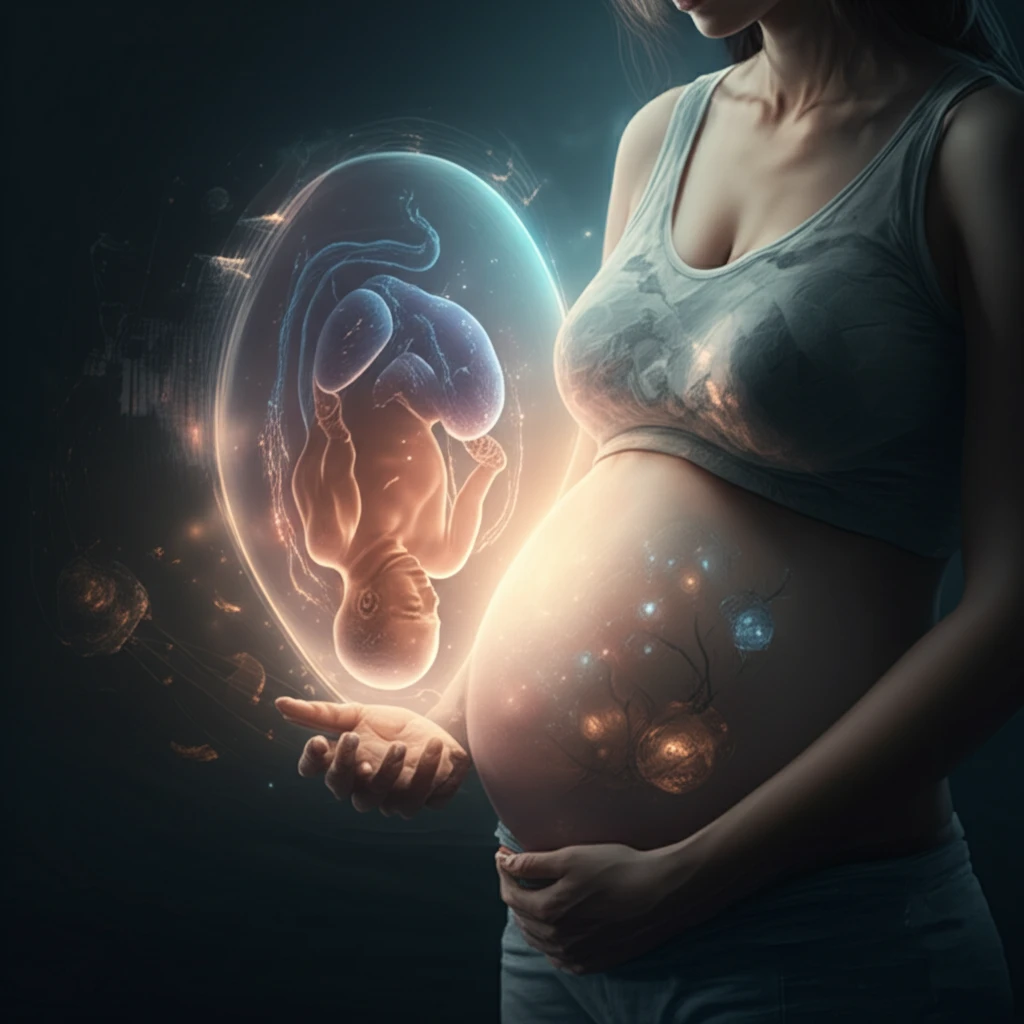
Traditionally, expectant parents rely on 2D ultrasound images to visualize their developing baby. However, these images can be difficult for non-medical professionals to interpret. 3D printing offers a tangible alternative, physically recreating the interior of the womb during gestation, showcasing the baby's physical appearance and actual size. In cases of malformation, these models can provide critical insights for medical planning and parental preparation.
Modern advancements in medical imaging, including scanning and automated image interpretation, play a vital role in healthcare by enabling earlier and more accurate diagnoses through non-invasive procedures. The integration of these imaging systems with additive manufacturing is proving to be a significant step forward. This combined approach spans a wide array of medical imaging modalities, leading to significant advancements, from tissue engineering to customized implants.
From Scans to Sculptures: How 3D Printing Works
The process begins with medical imaging techniques such as 3D ultrasound (3DUS), magnetic resonance imaging (MRI), and computed tomography (CT) scans. These technologies capture detailed images of the fetus within the womb, each offering unique advantages. Ultrasound is widely used due to its safety and cost-effectiveness, while MRI provides high-resolution images ideal for visualizing internal tissues. CT scans, though used less frequently due to radiation exposure, are valuable for examining skeletal structures.
- 3D Ultrasound (3DUS): Safe and cost-effective for routine visualization.
- Magnetic Resonance Imaging (MRI): High-resolution imaging of soft tissues.
- Computed Tomography (CT): Detailed imaging of skeletal structures.
- Specialized 3D Modeling Software: Converts medical images into printable models.
A New Dimension in Prenatal Understanding
The introduction of 3D printed fetal models marks a significant advancement in prenatal care. These models offer a novel approach to education, allowing medical students and expecting parents to gain a deeper understanding of fetal development. They also foster a stronger emotional connection between parents and their unborn child, transforming the abstract concept of a developing baby into a tangible reality. This innovation holds immense potential for enhancing the prenatal experience and improving outcomes for both parents and children.