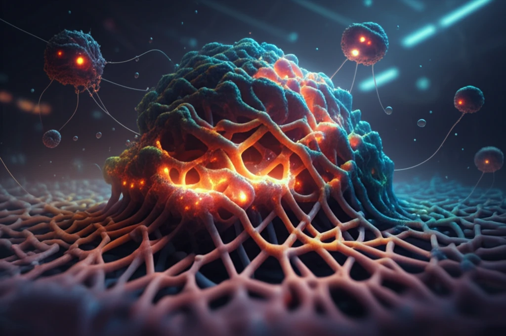
Intricate 3D-printed scaffolds supporting vibrant cell growth in a futuristic lab, representing tissue engineering advancements.

3D-Printed Cell Cultures: The Future of Tissue Engineering and Wound Healing?

"Explore how 3D-printed cell culture platforms are revolutionizing tissue engineering, offering unprecedented control over cell growth and structure for advanced medical applications."


For decades, the field of tissue engineering has strived to create functional human tissues outside the body. Essential to this endeavor are scaffolds—support structures that guide cell growth and organization. These scaffolds influence everything from cell adhesion and proliferation to differentiation, making their design crucial for successful tissue regeneration. Imagine a world where damaged organs can be repaired or replaced using tissues grown in the lab, all thanks to innovative scaffold technology.

Traditional approaches face limitations in replicating the complex environments cells naturally inhabit. This is where 3D printing steps in to change the game. By offering precise control over scaffold architecture, 3D printing enables scientists to create highly customized cell culture platforms. These platforms can be tailored to mimic the intricate structure of various tissues, providing an optimal environment for cell growth and tissue formation. For example, skin, being the body's protective barrier, is made of layers. 3D printing is creating innovative ways to regrow skin.

One promising 3D printing technique is electro-hydrodynamic jet (E-jet) printing, which uses an electric field to create fine jets of material. This method allows for the precise deposition of polymers like poly-(lactic-co-glycolic acid) (PLGA) to construct detailed 3D structures. Researchers are now exploring how varying parameters such as electrical voltage, plotting speed, and nozzle size affect the resulting cell culture platforms, aiming to optimize these platforms for specific tissue engineering applications.

The Science Behind 3D-Printed Cell Cultures

Intricate 3D-printed scaffolds supporting vibrant cell growth in a futuristic lab, representing tissue engineering advancements.

A recent study published in the Journal of Biomedical Materials Research delves into the use of E-jet 3D printing to create cell culture platforms for tissue engineering. The scientists focused on understanding how different printing parameters influence the structure and biocompatibility of PLGA scaffolds. By carefully controlling these parameters, they aimed to create platforms that promote cell adhesion, proliferation, and differentiation.

The researchers began by optimizing the E-jet printing process. They found that applying a high voltage to the nozzle (between 2.5 and 3.1 kV) created a stable jet suitable for printing. Higher voltages, however, resulted in unstable jets, underscoring the importance of fine-tuning the printing parameters. They also examined the effects of plotting speed and PLGA concentration, noting that both factors significantly impacted the structure of the printed filaments. For example, slower plotting speeds led to thicker fibers, while varying the PLGA concentration influenced the filament diameter.

The study's key findings include:
  • High voltage (2.5-3.1kV) allows best printing conditions.
  • Plotting speed matters in the thickness of fibers.
  • PLGA concentration effects filament diameter.
  • Smaller nozzle helps to reduce filament diamter.
To assess the biocompatibility of the 3D-printed scaffolds, the researchers cultured fibroblasts—the main cellular components of connective tissue—on the platforms. They found that the E-jet printed platforms supported cell adhesion and proliferation. Moreover, when the platforms were designed with aligned fibers, the fibroblasts exhibited a consistent orientation, suggesting that the scaffolds could guide cell alignment and growth. This alignment is crucial for creating functional tissues with specific mechanical properties.

The Future of Tissue Engineering

This research highlights the enormous potential of 3D-printed cell cultures in tissue engineering and regenerative medicine. By providing precise control over the cellular environment, these platforms offer a pathway to create functional tissues for a variety of applications, from wound healing to organ repair. As the technology advances, we can anticipate even more sophisticated designs that mimic the complexity of human tissues, paving the way for transformative medical breakthroughs.

About this Article -

This article was crafted using a human-AI hybrid and collaborative approach. AI assisted our team with initial drafting, research insights, identifying key questions, and image generation. Our human editors guided topic selection, defined the angle, structured the content, ensured factual accuracy and relevance, refined the tone, and conducted thorough editing to deliver helpful, high-quality information.See our About page for more information.

This article is based on research published under:

DOI-LINK: 10.1002/jbm.a.36188, Alternate LINK

Title: Control Of Cell Growth On 3D-Printed Cell Culture Platforms For Tissue Engineering

Subject: Metals and Alloys

Journal: Journal of Biomedical Materials Research Part A

Publisher: Wiley

Authors: Zhikai Tan, Tong Liu, Juchang Zhong, Yikun Yang, Weihong Tan

Published: 2017-09-15

Everything You Need To Know

1

How are 3D-printed cell culture platforms revolutionizing tissue engineering, and what advantages do they offer over traditional methods?

3D-printed cell culture platforms revolutionize tissue engineering by offering unprecedented control over cell growth and structure. This is achieved through the use of scaffolds, which are support structures that guide cell adhesion, proliferation, and differentiation. These platforms can be tailored to mimic the intricate structure of various tissues, providing an optimal environment for tissue formation. This approach contrasts with traditional methods that face limitations in replicating the complex environments cells naturally inhabit. The advancement is particularly impactful in creating skin, the body's protective barrier, through innovative regeneration methods.

2

What is E-jet printing, and how do parameters like electrical voltage and plotting speed influence the construction of 3D structures using materials such as PLGA?

E-jet printing utilizes an electric field to create fine jets of materials, allowing precise deposition of polymers such as poly-(lactic-co-glycolic acid) or PLGA, to construct detailed 3D structures. Key parameters, including electrical voltage, plotting speed, and nozzle size, affect the resulting cell culture platforms. For instance, high voltage (2.5-3.1kV) allows best printing conditions, plotting speed influences fiber thickness, PLGA concentration affects filament diameter, and smaller nozzles reduce filament diameter. The optimization of these parameters is critical for specific tissue engineering applications, influencing the structure and biocompatibility of PLGA scaffolds.

3

How does the concentration of PLGA impact the structure of 3D-printed cell culture platforms, and why is this important in tissue engineering?

PLGA, or poly-(lactic-co-glycolic acid), is used in E-jet printing to create 3D structures for cell culture platforms. Its concentration is a key factor influencing the filament diameter of these structures. Researchers manipulate PLGA concentration, along with other parameters such as electrical voltage and plotting speed, to optimize the cell culture platforms. This optimization is crucial for promoting cell adhesion, proliferation, and differentiation, essential for successful tissue engineering.

4

What role do fibroblasts play in assessing the biocompatibility of 3D-printed scaffolds, and how does the alignment of fibers in these scaffolds affect cell behavior?

Fibroblasts, the main cellular components of connective tissue, are cultured on 3D-printed scaffolds to assess their biocompatibility. These scaffolds support cell adhesion and proliferation, and when designed with aligned fibers, induce fibroblasts to exhibit a consistent orientation. This alignment is crucial for creating functional tissues with specific mechanical properties, indicating that the scaffolds can guide cell alignment and growth, essential for tissue regeneration and functionality.

5

What are the future implications of using 3D-printed cell cultures in regenerative medicine, and how might this technology lead to transformative medical breakthroughs beyond wound healing?

3D-printed cell cultures hold immense potential in tissue engineering and regenerative medicine by providing precise control over the cellular environment. This precision allows for the creation of functional tissues for wound healing and even organ repair. As the technology advances, more sophisticated designs mimicking the complexity of human tissues are anticipated. The ability to control cell alignment and growth using materials like PLGA, optimized through parameters such as voltage and plotting speed in E-jet printing, paves the way for transformative medical breakthroughs. Though not discussed here, the technology could extend to personalized medicine where scaffolds are tailored to an individual's own cells, enhancing compatibility and reducing rejection risks.

Newsletter Subscribe

Subscribe to get the latest articles and insights directly in your inbox.