
Knee Deep in Innovation: How MRI and Magnetic Stem Cell Targeting Are Revolutionizing Cartilage Repair

"Discover how a groundbreaking study is using MRI to assess and improve magnetic stem cell therapy for damaged knees, offering hope for athletes and arthritis sufferers."
Knee pain, a familiar woe to athletes and those battling arthritis, often stems from cartilage damage. This resilient tissue, essential for smooth joint movement, lacks the inherent ability to heal itself due to its limited blood supply. As a result, even minor injuries can lead to chronic pain and reduced mobility. But, traditional treatments often involve invasive surgeries and extended recovery periods.
Fortunately, regenerative medicine offers a beacon of hope, particularly through stem cell therapies. Scientists have explored various methods of cartilage repair, with mesenchymal stem cells (MSCs) taking center stage. These cells, capable of differentiating into cartilage-producing cells, can be transplanted into damaged joints to stimulate healing. However, delivering MSCs effectively and ensuring their retention at the injury site has been a significant challenge.
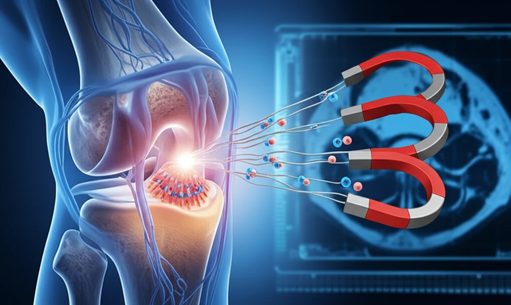
Now, imagine if we could guide stem cells directly to the damaged area using magnets. Recent research has made this a reality. A pioneering approach called magnetic stem cell targeting uses magnetic fields to deliver MSCs precisely to cartilage defects. A new study investigates the effectiveness of this method, combined with the power of magnetic resonance imaging (MRI) to monitor the repair process. The results are promising, offering new hope for those seeking less invasive and more effective cartilage repair options.
The Scale of Cartilage Repair Innovation
Cartilage repair is an increasingly active area of clinical development. ChondroFiller, an injectable collagen scaffold, has been shown to improve functional IKDC scores from 48 to 80 over three years in independent European cohorts by recruiting the body's own progenitor cells without surgery or general anaesthetic. Minced cartilage implantation has also become an increasingly utilized technique, though notable variations remain in how the procedure is performed across centers. Researchers at Duke Health have found that the ankle's ability to regenerate cartilage shares mechanisms with limb regeneration in animals such as salamanders and zebrafish, pointing toward broader therapeutic possibilities for osteoarthritis.
Established Techniques and Their Shortcomings
Cartilage tissue engineering has long been limited by articular cartilage's inherently poor capacity for self-repair, with early attempts relying on injecting cultured autologous chondrocytes under a periosteal flap. Current review literature describes three broad approaches at the rheumatology–orthopaedics interface: disease-modifying osteoarthritis drug (DMOAD) treatment, cell-based therapies, and intrinsic cartilage repair through joint distraction. Established techniques such as autologous chondrocyte implantation (ACI) demonstrate 10–17 year durability for focal knee defects, yet mesenchymal stem cell therapies have not been rigorously tested against them in large-scale comparative trials, leaving a significant evidence gap.
From Biological Foundations to Modern Milestones
Hyaline cartilage—composed primarily of water, collagen fibers, and chondrocytes—provides the smooth, resilient surface that enables comfortable joint movement, yet it possesses a limited capacity for self-repair. Significant milestones have since reshaped clinical practice: the STACi technique now condenses what once required two operations spanning three to six months into a single operative session lasting 2.5 to 4 hours. Research into so-called 'dancing molecules' has also gained attention, with studies suggesting these dynamic structures may be particularly effective at stimulating cartilage repair in osteoarthritic joints.
Magnetic Precision Meets Imaging Clarity
The study, published in Tissue Engineering, delves into the use of MRI to evaluate cartilage repair following magnetic delivery of stem cells. Researchers at Hiroshima University in Japan conducted a detailed experiment using rabbits to assess this innovative approach. They induced articular cartilage defects in the rabbits' knees and then treated them with magnetically labeled bone marrow mesenchymal stem cells (MSCs).
- T2 mapping of MRI successfully reflected the degree of cartilage repair, with the m-MSC group showing significantly better repair at 12 weeks.
- T2 images revealed hypointense areas in the m-MSC group, indicating the presence of iron particles from the magnetically labeled MSCs.
- Histological analysis confirmed the presence of iron particles in the repaired tissues at 1 and 4 weeks, but not at 12 weeks, suggesting their gradual metabolism.
Emerging Therapies and Key Distinctions
Injectable hydrogels represent a major frontier in cartilage tissue engineering, with recent reviews thoroughly cataloging the polymers, cells, and stimulators commonly used in their preparation alongside future challenges for clinical translation. A key conceptual advance emphasized in current stem cell research is the distinction between cartilage repair—which produces tissue structurally similar to hyaline cartilage at the macroscopic level—and true cartilage regeneration, which is an entirely different biological process. Contrary to longstanding belief, Duke Health researchers have reported that cartilage in human joints can repair itself through a process similar to that used by salamanders and zebrafish to regenerate limbs.
Clinical Limitations and Early Failures
Despite decades of progress, tissue engineering for articular cartilage repair continues to face practical clinical limitations, particularly surrounding the use of autologous adult cells. Early clinical failures with scaffold plugs designed for cartilage repair in the knee have been documented, underscoring the gap between laboratory promise and real-world outcomes. The field has demonstrated that cartilage tissue engineering is possible in principle, but translating that principle into reliable, widely reproducible clinical results remains a persistent challenge.
Market Landscape and Procedural Trade-Offs
The global cartilage repair market was valued at USD 1.7 billion in 2026 and is projected to grow at a compound annual growth rate of 5.8%, reaching an estimated USD 3.0 billion by 2036, with Johnson & Johnson, Smith+Nephew, Stryker, Arthrex, and Zimmer Biomet identified as major participants. Academic analyses have proposed that the ideal future procedure for articular cartilage repair will combine the ease of one-step microfracture with the chondrogenic benefits of ACI while mitigating each method's respective limitations. This vision of a hybrid approach reflects the growing consensus that no single existing technique fully addresses all clinical needs.
The Future of Knee Care is Looking Up
This research marks a significant step forward in the field of regenerative medicine. By combining magnetic stem cell targeting with MRI evaluation, scientists have developed a powerful tool for assessing and improving cartilage repair. This innovative approach holds great promise for treating knee injuries and arthritis, offering patients less invasive and more effective treatment options. As research progresses, we can anticipate further refinements and broader applications of this technology, ultimately leading to improved outcomes and a better quality of life for those suffering from joint damage.
Expert Perspectives on Current Therapeutics
Systematic analyses have evaluated the clinical efficacy of microfracture for articular cartilage repair in the knee, forming a key part of the evidence base for current therapeutic guidelines. However, an expert opinion published in Expert Opinion on Biological Therapy concluded that, to date, there is no established way to derive a chondrogenic lineage from stem cells that forms functional hyaline cartilage tissue in vivo. One proposed alternative is to enhance the chondrogenic potential of repair cells already present in diseased cartilage tissue rather than relying on externally introduced stem cells. Articular cartilage remains an avascular tissue with limited inherent ability to repair and self-renew, a fundamental constraint that continues to shape therapeutic strategy.
Market Growth and Projected Trajectories
The orthopedic cartilage repair market is expected to sustain meaningful growth through 2033, reflecting increasing clinical adoption and ongoing research investment. More specifically, the cartilage repair devices segment is projected to reach USD 17.23 billion by 2035, expanding at a compound annual growth rate of 18.5% according to Market Research Future. These projections suggest that technological refinements, broader clinical evidence, and rising patient demand will continue to drive the market's upward trajectory over the coming decade.
Structural Hurdles in Cartilage Repair
While scientific advances in cartilage repair have been substantial, the field continues to grapple with systemic challenges that no single breakthrough has yet resolved. Access to advanced therapies remains uneven across healthcare systems, and the high cost of procedures such as ACI limits their availability to well-resourced clinical settings. Regulatory pathways for novel biologics and cell-based treatments add further complexity, often prolonging the time between laboratory discovery and widespread patient access. Addressing these broader structural barriers will be essential to translating scientific progress into meaningful, equitable improvements in patient outcomes.
Translating Innovation into Patient Outcomes
A 2025 feasibility study using ChondroFiller in the wrist confirmed superior Outerbridge and ICRS cartilage scores at follow-up, though that study employed a surgical delivery route distinct from the ultrasound-guided outpatient injection pathway. Clinical studies have also demonstrated improved cartilage regeneration following biomaterial implantation after bone marrow stimulation, reinforcing the value of scaffold-based approaches in regenerative joint surgery. Yet for patients with severe cartilage damage, stem cell therapies do not rebuild cartilage from scratch, and joint replacement remains the only option that fully restores function—though it is irreversible. This reality ensures that less invasive regenerative strategies remain one of the most intensely studied fields in orthopedics.