3D Bioprinting: Revolutionizing Breast Cancer Drug Resistance Studies
"Discover how scientists are using 3D bioprinting to create more realistic breast cancer models for better drug screening and personalized medicine."
Breast cancer is a complex disease influenced by its surrounding environment, known as the microenvironment. This microenvironment includes various cells like immune cells, endothelial cells, cancer-associated fibroblasts (CAFs), and adipose-derived mesenchymal stem cells (ADMSCs). Understanding how these components interact is crucial for developing effective treatments.
Traditional two-dimensional (2D) cell cultures have limitations in replicating the complexity of the in vivo tumor environment. As a result, three-dimensional (3D) models are gaining traction, offering a more accurate representation of cell-cell and cell-matrix interactions. Among these, 3D bioprinting stands out as a promising technique to assemble living cells and biomaterials into custom-designed constructs.
Recent research published in ACS Biomaterials Science & Engineering explores the use of 3D bioprinting to create breast cancer models for drug resistance studies. This innovative approach allows scientists to investigate the effects of ADMSCs on breast cancer cells in a more realistic setting, potentially leading to better drug screening and personalized medicine strategies.
3D Bioprinting Breast Cancer Models: A Step-by-Step Look
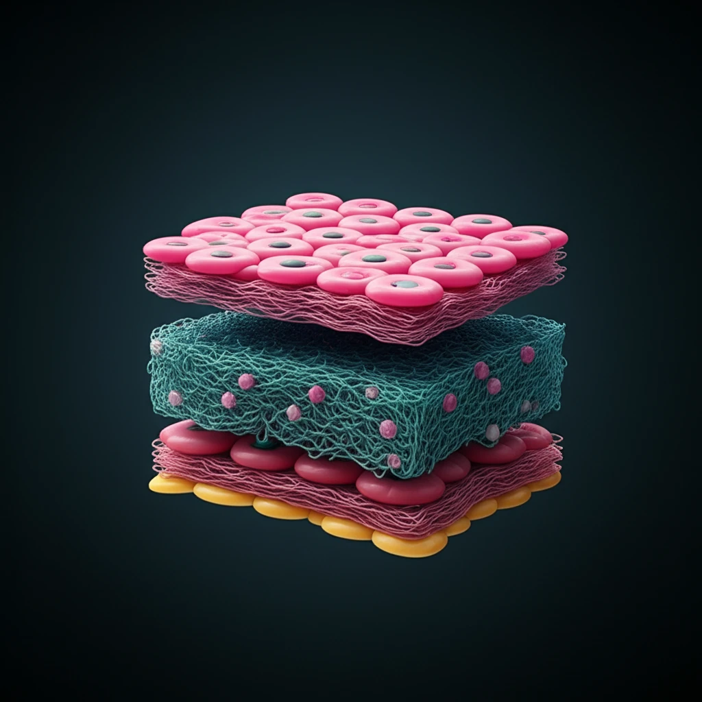
The study utilized 3D bioprinting to construct models with breast cancer cells (21PT, HER2+) in the middle and ADMSCs surrounding them, mimicking the in vivo structure of a tumor. These constructs were created using dual hydrogel-based bioinks, allowing for precise placement of different cell types.
- Reduced Apoptosis: The percentage of cleaved Caspase-3 positive cells, indicating apoptosis (cell death), was significantly lower in the 3D bioprinted constructs with ADMSCs compared to cancer cells alone, especially at low DOX doses.
- ADMSC Thickness Matters: Increasing the thickness of the ADMSC layer further enhanced drug resistance, suggesting that ADMSCs provide a protective effect to cancer cells.
- LOX Involvement: Both ADMSCs and 21PT cells expressed lysyl oxidase (LOX), an enzyme involved in collagen cross-linking and known to play a role in cancer progression. Inhibition of LOX enhanced DOX sensitivity.
The Future of Cancer Research: 3D Bioprinting and Personalized Medicine
This study underscores the potential of 3D bioprinting to revolutionize cancer research by creating more realistic and predictive tumor models. By incorporating key components of the tumor microenvironment, such as ADMSCs, these models can better mimic the complex interactions that influence drug response.
Furthermore, the ability to control parameters like ADMSC layer thickness allows researchers to investigate the impact of obesity and other microenvironmental factors on drug resistance. The identification of LOX as a potential target to enhance drug sensitivity opens new avenues for therapeutic intervention.
As 3D bioprinting technology advances, it promises to play an increasingly important role in personalized medicine, enabling the development of tailored cancer treatments based on the unique characteristics of individual tumors and their microenvironment.