
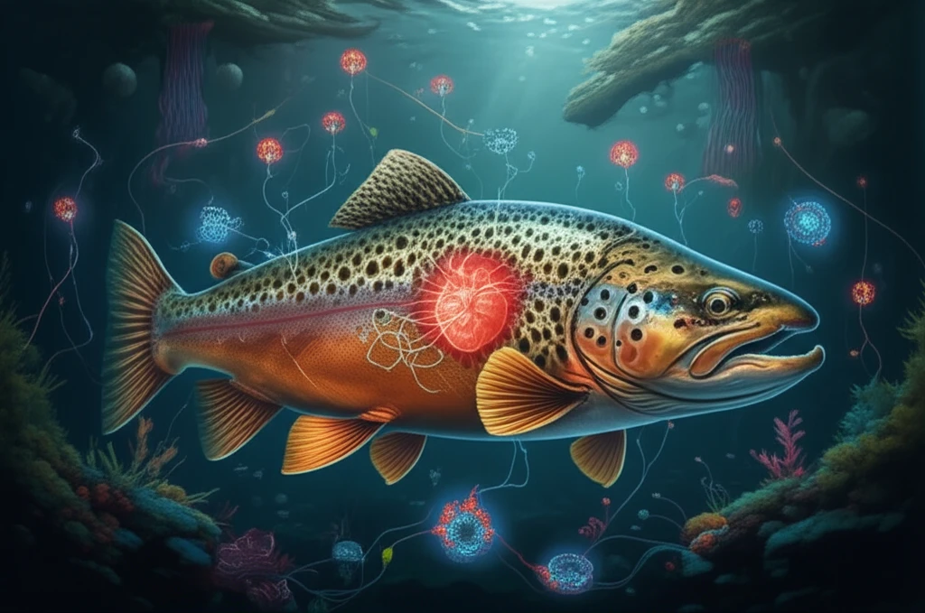
Why Your Heart Beats a Little Differently: Unraveling the Mysteries of Fish Pacemakers

"Scientists discover unique features in trout hearts that challenge what we know about cardiac rhythms and could offer new insights into heart health."
Our hearts, those tireless engines within us, beat to a rhythm orchestrated by a complex interplay of biological mechanisms. Central to this rhythm is the sinoatrial (SA) node, often dubbed the heart's natural pacemaker. Within this node, specialized cells generate electrical impulses that spread throughout the heart, triggering the coordinated contractions that pump blood through our bodies. The rate at which these impulses fire—our heart rate—is exquisitely sensitive to a variety of factors, from our body temperature and activity level to our emotional state.
For decades, scientists have believed that a particular type of ion channel, responsible for the 'funny current' (If), plays a crucial role in regulating the heart's pacemaker function. These channels, activated by hyperpolarization (a decrease in voltage across the cell membrane), were thought to be essential for the rhythmic firing of SA node cells. However, recent research into the hearts of brown trout (Salmo trutta fario) is challenging this long-held assumption, revealing a surprising degree of diversity in cardiac pacemaking mechanisms across the animal kingdom.
In fishes, heart rate is supremely adaptable, responding swiftly to changes in water temperature. This is critical for their survival, as temperature directly affects their metabolic rate and oxygen demands. Understanding how fish hearts regulate their rhythm, therefore, offers valuable insights into the fundamental limits of physiological adaptation. This study could also explain variations in effectiveness of cardiac drugs.
The Fish Heart as a Window into Cardiac Pacemaking
The fish heart serves as a crucial model for understanding cardiac pacemaking due to its conserved physiological mechanisms. Research highlights the significance of T-type Ca2+ channels in temperature-related modulation of fish cardiac pacemaking and impulse conduction. Molecular and cellular processes regulating cardiac excitation-contraction coupling influence the types of cardiac arrhythmias triggered by natural and anthropogenic environmental factors in fish. The regulation of electrical excitability of cardiac myocytes in the pacemaker region, atrium, and ventricle is a key factor determining the pumping function of the fish heart.
Common Research Methods in Fish Cardiac Studies
Researchers typically employ electrocardiography and optical mapping techniques to study fish cardiac pacemaking, though these methods have inherent limitations in capturing the full complexity of cardiac function. Invasive approaches often require immobilization or anesthesia, which can alter normal cardiac physiology. The small size of many fish hearts presents technical challenges for high-resolution recording and manipulation. Additionally, translating findings from ectothermic fish to endothermic mammals remains a significant methodological hurdle.
From Galvani to Modern Pacemaking Science
The history of cardiac pacemaking research spans over two centuries, beginning with Luigi Galvani's discovery of the heart's response to electrical currents in the late 18th century. Paul Zoll's pioneering work in the 1950s introduced external pacemakers and defibrillators, marking a revolutionary milestone in cardiac medicine. Cardiac pacemaking has been thoroughly investigated for decades using a multiscale approach at organ, tissue, cell, and molecular levels to clarify the basic mechanisms underlying rhythm generation and control. Even in simple contractile cardiac tubes in primitive invertebrates, cardiac function is controlled by intrinsic, autonomous pacemaker cells, demonstrating deep evolutionary conservation.
Trout Hearts: Beating to a Different Drummer?
Researchers at the University of Eastern Finland embarked on a detailed investigation of brown trout hearts, seeking to understand the role of If channels in their cardiac pacemaking. What they discovered was quite unexpected. While trout hearts possessed the genetic machinery to produce If channels—encoding six different HCN channel transcripts (HCN1, HCN2a, HCN2ba, HCN2bb, HCN3, and HCN4)—the actual contribution of these channels to the heart's rhythm appeared to be minimal.
- Low If Current Density: Only a small percentage of sinoatrial pacemaker cells exhibited a measurable If current, and even when present, the current density was remarkably low compared to mammals.
- Limited Impact of If Blockade: Blocking If channels with cesium (Cs+) had only a modest effect on the heart rate of excised trout heart tissue.
- Alternative Mechanisms: The team found SR Ca2+ management to be playing larger role than If Channels.
Zebrafish Cardiac Regeneration Breakthroughs
Zebrafish have emerged as a key model organism for cardiac regeneration research due to their extraordinary regenerative capabilities, particularly in the heart. Recent findings challenge the long-held belief that oxidative phosphorylation is detrimental for heart regeneration, as it was thought reactive oxygen species block cardiomyocyte proliferation through DNA damage. The zebrafish's transparency, genetic tractability, and conserved cardiac physiology make it a powerful vertebrate model for cardiovascular research. Understanding how signaling pathways link the cardiac endothelium to heart regeneration offers new therapeutic possibilities.
Challenges in Fish Pacemaking Research
Despite advances, significant gaps remain in understanding fish cardiac pacemaking mechanisms, particularly regarding species-specific variations across the approximately 30,000 fish species. The molecular basis of fish cardiac T-type calcium channels remains poorly characterized, limiting comprehensive mechanistic understanding. Translating fish cardiac research to human clinical applications faces substantial hurdles due to fundamental physiological differences between ectothermic and endothermic species. Additionally, standardizing experimental conditions across diverse aquatic environments presents ongoing methodological challenges.
Cross-Species Perspectives on Cardiac Function
Comparative studies reveal that fish possess the greatest range for central arterial blood pressure and relative ventricular mass of any vertebrate group, demonstrating remarkable cardiac diversity. This enormous variation provides excellent material for mechanistic studies linking organ form and function with their molecular underpinnings. Fish cardiac myocytes rely more heavily on cycling of extracellular calcium rather than intracellular calcium stores from the sarcoplasmic reticulum for contraction activation. Such comparative insights illuminate fundamental principles of cardiac evolution and adaptation.
Implications for Heart Health and Beyond
The discovery of a novel cardiac pacemaking mechanism in brown trout raises important questions about the diversity of heart function across the animal kingdom. It suggests that the reliance on If channels may be a relatively recent evolutionary development, or that fish have evolved alternative strategies to achieve the same physiological outcome. Further research into the hearts of other fish species will be needed to determine how widespread this phenomenon is.
Integrative Autonomic Control in Fish Hearts
The isolated, innervated zebrafish heart has been established as a novel alternative model for studies of autonomic control of heart rate. Stimulation of individual cardiac vagosympathetic nerve trunks evoked both bradycardia through parasympathetic activation and tachycardia through sympathetic activation. Ultrastructure, molecular composition, and electrophysiological properties of fish cardiac myocytes suggest that cycling of extracellular calcium is generally more important than intracellular cycling of calcium stores in activating contraction. These findings demonstrate the conserved nature of autonomic cardiac regulation across vertebrate evolution.
Emerging Directions in Fish Cardiac Research
Future research will likely focus on unraveling the complete molecular mechanisms underlying fish cardiac pacemaking, particularly the role of T-type calcium channels in temperature modulation. Advanced imaging technologies and genetic tools will enable higher resolution studies of cardiac function in vivo. Integration of multi-omics approaches promises to reveal new regulatory networks controlling cardiac rhythm. These advancements may ultimately inform therapeutic strategies for human cardiac arrhythmias.
Biohybrid Technologies and Extreme Environment Adaptations
Researchers have developed biohybrid fish made from human cardiac cells that swim through the coordinated contraction of cardiac muscle cells. This technology builds on previous work using rat cardiac cells to create jellyfish-like biohybrid pumps and swimming artificial stingrays. Understanding how fish maintain cardiac pacemaking in extreme conditions provides insights into the limits of biological adaptation. Cardiac performance is tightly related to heart rate, and extrinsic and intrinsic control of pacemaker activity has been explored to understand rhythm modulation under extreme environments.
Clinical Translational Potential
Natural selection has produced many vertebrate solutions for the cardiac life-support system, especially among approximately 30,000 fish species, offering a vast reservoir of biological insights. The enormous cardiac diversity among fish provides excellent ground material for mechanistic studies linking form and function. Clinical studies like FISH&CHIPS have demonstrated the positive impact of advanced cardiac imaging analysis for diagnosing coronary artery disease. Real-world implementation of such technologies in healthcare systems shows significant potential for reducing cardiovascular mortality.