
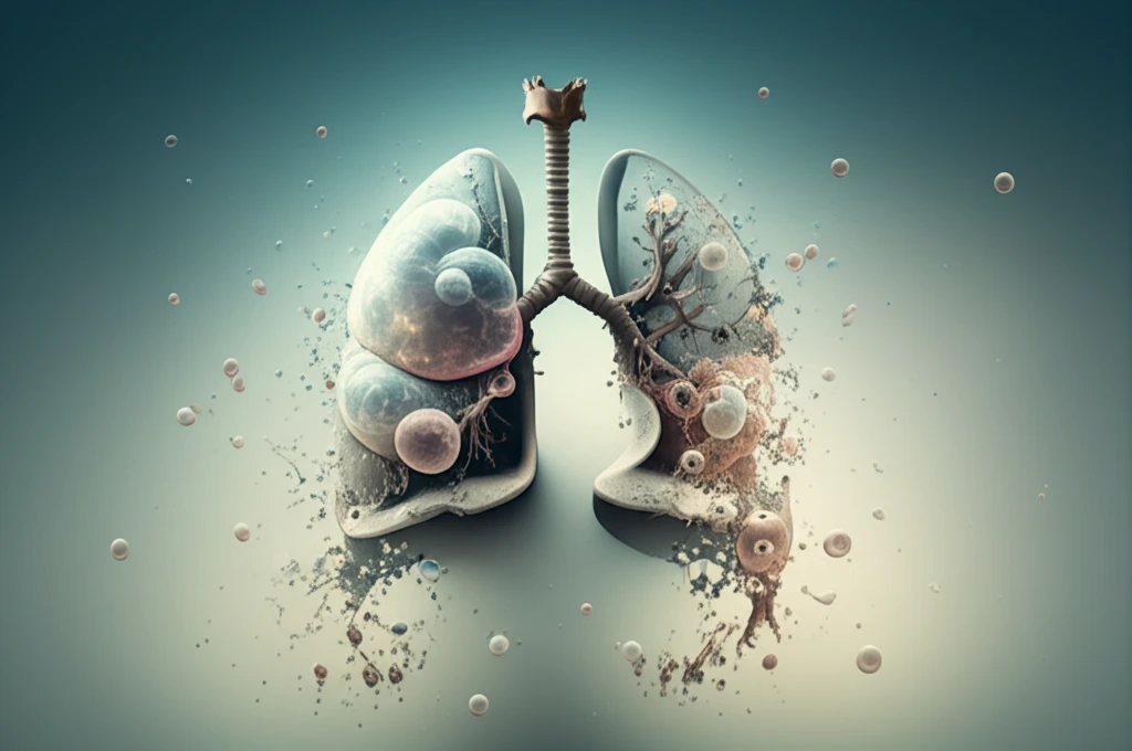
Vanishing Lung Syndrome: What You Need to Know About This Rare Condition

"Understanding the causes, symptoms, and treatment options for vanishing lung syndrome to ensure early diagnosis and better management."
Imagine struggling to breathe, feeling as though your lungs are gradually disappearing. This is the reality for individuals with Vanishing Lung Syndrome (VLS), a rare and progressive condition that primarily affects smokers. Also known as giant bullous emphysema (GBE), VLS is characterized by the formation of large air-filled sacs (bullae) in the lungs, which can compress healthy tissue and impair respiratory function.
The term 'vanishing lung' might sound alarming, but understanding the condition is the first step toward managing it. This article aims to provide a comprehensive overview of VLS, covering its causes, symptoms, diagnosis, and available treatment options. Whether you're a healthcare professional, a patient, or simply interested in learning more, this guide will offer valuable insights into this rare pulmonary disorder.
While VLS is often associated with long-term smoking, it's important to recognize that not everyone who smokes will develop this condition. Genetic factors and other environmental exposures may also play a role. Early diagnosis and appropriate management can significantly improve the quality of life for those affected, making awareness and education crucial.
Defining VLS and Its Clinical Burden
Vanishing lung syndrome (VLS), also known as idiopathic giant bullous emphysema, is defined by the emergence of sizable bullae causing compression on healthy lung tissue. Diagnosis is based on radiographic evidence showcasing giant bullae occupying at least one-third of the hemithorax in one or both lungs. The condition is considered rare, and treatment options include lung-volume-reduction surgery for selected patients after thorough assessment. Precise prevalence data remain limited, reflecting the syndrome's rarity and the challenges of systematic epidemiological study.
Current Diagnostic and Therapeutic Approaches
Diagnosis of vanishing lung syndrome relies primarily on imaging, including chest X-ray and high-resolution CT scans, to identify giant bullae compressing normal lung parenchyma. Treatment ranges from conservative management—such as smoking cessation and pulmonary rehabilitation—to surgical interventions including bullectomy and lung-volume-reduction surgery for advanced cases. However, detailed comparative effectiveness data for these approaches remain sparse, and treatment decisions are often guided by individualized clinical judgment rather than large-scale randomized trials. The rarity of the condition limits the development of standardized, evidence-based treatment protocols.
Early Descriptions and Defining Criteria
Vanishing lung syndrome was first described in the medical literature as a case of vanishing lungs with multiple giant bullae occupying both hemithoraces. Roberts and colleagues subsequently established the imaging criteria defining VLS as giant bullae involving at least one-third of one or both lungs while compressing surrounding normal lung parenchyma on radiographs. The condition is also classified as Type I bullous disease, or primary bullous disease of the lung, and has historically been most commonly identified in young male smokers. These foundational definitions have remained central to diagnosis and classification of VLS in subsequent decades.
What Causes Vanishing Lung Syndrome?
Vanishing Lung Syndrome is most commonly linked to chronic obstructive pulmonary disease (COPD), which is frequently caused by long-term smoking. The persistent inflammation and damage to the lung tissue lead to the formation of bullae. These bullae are essentially enlarged airspaces that don't contribute to oxygen exchange and can compress the surrounding healthy lung tissue.
- Alpha-1 Antitrypsin Deficiency: A genetic condition that can lead to early-onset emphysema and bullae formation.
- Environmental Exposures: Prolonged exposure to pollutants, dust, and fumes can irritate the lungs and increase the risk of COPD and VLS.
- Other Lung Diseases: Conditions like asthma and cystic fibrosis can sometimes lead to the development of bullae.
Contemporary Case Reports and Systematic Reviews
Recent literature, including a 2024 systematic review, continues to characterize VLS as a condition where bullae impair ventilation because they do not participate in gas exchange, leading to worsening dyspnea that often necessitates surgical intervention. A case report of a 36-year-old patient highlighted that VLS can present even in younger individuals without extensive smoking histories, broadening the previously understood demographic profile. The systematic review component of this work aimed to discern demographics, risk factors, and treatment modalities across the existing body of VLS literature. The elusive etiology of VLS continues to challenge researchers, with diagnosis still fundamentally dependent on radiographic evidence.
Limitations of Surgical Intervention
Lung-volume-reduction surgery, while considered for selected VLS patients, is not universally effective and requires careful patient selection based on exercise capacity, pulmonary-function testing, and confirmed smoking cessation. Not all patients are suitable surgical candidates, and the procedure carries inherent risks that must be weighed against potential benefits. The requirement for smoking cessation before surgery highlights a significant barrier, as many VLS patients continue tobacco use despite diagnosis. These constraints underscore that surgical intervention, though a key therapeutic option, has notable limitations and failure points that must be acknowledged in clinical practice.
VLS in the Broader Landscape of Bullous Lung Disease
Vanishing lung syndrome occupies a distinct position within the spectrum of bullous lung diseases, differentiated by the sheer scale of bullae involvement—at least one-third of the hemithorax. While related to chronic obstructive pulmonary disease and other forms of emphysema, VLS is distinguished by its radiographic presentation and the progressive compression of otherwise healthy lung tissue. The rarity of VLS compared to more common pulmonary conditions means that direct comparative studies are scarce, and clinicians often draw on broader emphysema research to inform management decisions. Further research is needed to clarify how VLS outcomes compare with those of other bullous and emphysematous conditions.
Living with Vanishing Lung Syndrome: A Focus on Quality of Life
While VLS presents significant challenges, advancements in treatment and management strategies offer hope for improved quality of life. By understanding the condition, seeking early diagnosis, and adhering to a comprehensive treatment plan, individuals with VLS can better manage their symptoms and live more fulfilling lives. Continuous research and innovation in pulmonary medicine are paving the way for even more effective therapies in the future.
Expert Perspectives on VLS Management
Clinical experts broadly agree that early identification of vanishing lung syndrome through imaging is critical to preventing irreversible lung damage and deteriorating respiratory function. The consensus view emphasizes a multidisciplinary approach combining imaging expertise, pulmonology, and thoracic surgery to optimize patient outcomes. Expert commentary in recent literature stresses the importance of addressing modifiable risk factors, particularly smoking, as a cornerstone of both prevention and treatment. While no universally accepted treatment algorithm exists, the integration of individualized care plans remains the prevailing expert recommendation.
Emerging Questions and Research Directions
The elusive etiology of vanishing lung syndrome remains a significant gap in current knowledge, with research ongoing to better understand the underlying mechanisms driving giant bullae formation. Advances in imaging technology may improve early detection and allow for more precise characterization of disease progression. Future studies are expected to focus on larger patient cohorts and potentially randomized comparisons of surgical versus conservative management strategies. The development of targeted therapies, if the molecular basis of VLS becomes clearer, represents a longer-term frontier that could transform treatment paradigms.
Challenges in Addressing a Rare Condition
Vanishing lung syndrome faces systemic challenges common to rare diseases, including limited awareness among general practitioners and potential delays in diagnosis. The small patient population makes it difficult to conduct large-scale clinical trials, leaving treatment decisions heavily reliant on case reports and expert opinion. Resource allocation for research into rare pulmonary conditions is inherently constrained compared to more prevalent diseases such as standard COPD or lung cancer. Addressing these systemic barriers will require coordinated efforts across research institutions, funding bodies, and clinical networks to build the evidence base needed for improved patient care.
Patient Experiences and Quality of Life
Patients with vanishing lung syndrome often endure a long history of cigarette smoking and/or marijuana use alongside chronic obstructive pulmonary disease, contributing to significant declines in quality of life. Case reports, such as that of a 36-year-old female smoker, illustrate that VLS can affect individuals at relatively young ages and profoundly disrupt daily functioning. The progressive nature of the condition means patients frequently experience worsening dyspnea that limits physical activity and independence. Real-world patient outcomes underscore the human toll of VLS and the urgent need for both prevention strategies and effective therapeutic interventions.