
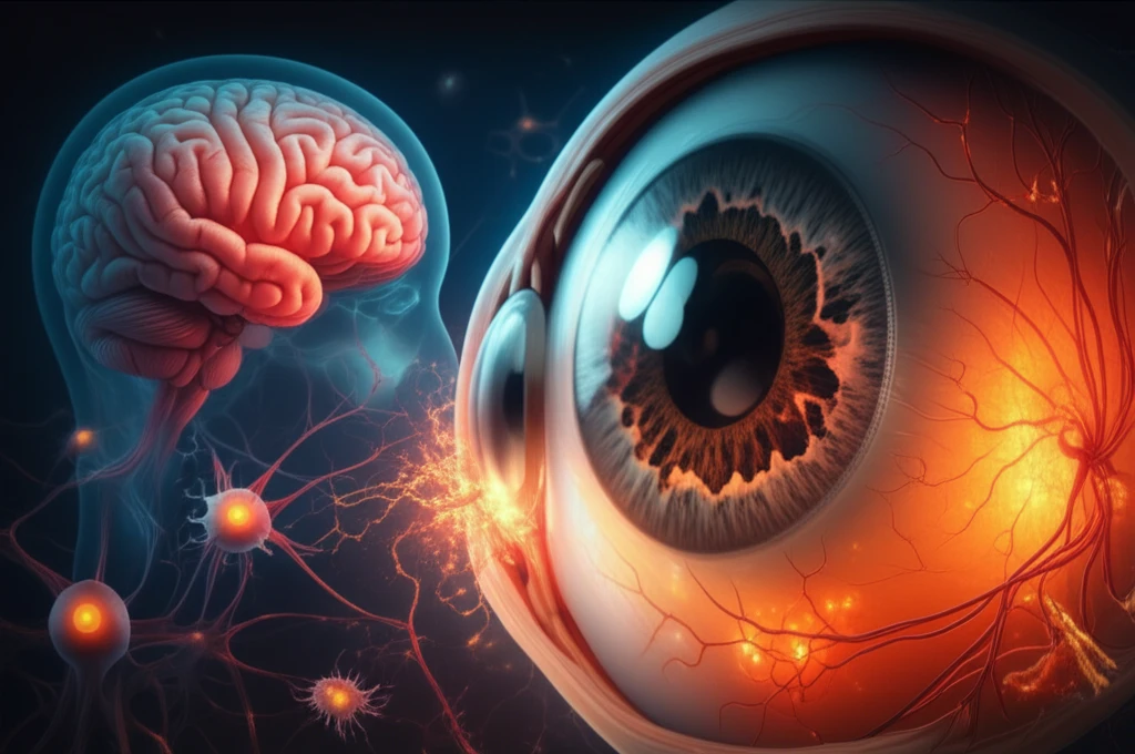
Illustration depicting the PVL toxin attacking the human eye and brain, highlighting the impact on retinal cells and neurons.

Unveiling the Enemy Within: How a Common Bacterial Toxin Targets Your Brain and Eyes

"Groundbreaking research reveals how a toxin from Staphylococcus aureus directly attacks brain and eye cells, offering new insights into infection and potential treatments."


In the microscopic world, a silent war is constantly being waged within our bodies. Bacteria, often unseen, can unleash potent toxins that wage war against our cells. One such toxin, Panton-Valentine Leukocidin (PVL), produced by the common bacterium Staphylococcus aureus (S. aureus), has recently come under the spotlight for its surprising and devastating effects on our bodies. This article dives deep into cutting-edge research that reveals how PVL directly targets brain and eye cells, offering a new understanding of infection and paving the way for potential treatments.

S. aureus, often found on the skin and in the nasal passages of healthy individuals, can turn into a formidable foe when it produces PVL. This toxin is particularly dangerous because it attacks and destroys white blood cells, weakening the body's defenses and leading to severe infections. However, the story doesn't end there. Recent studies have shown that PVL's reach extends far beyond the typical infection sites, with its toxic effects now linked to the brain and eyes.

This groundbreaking research, detailed in a recent study, used a rabbit retinal explant model to investigate the effects of PVL on the delicate tissues of the eye. The findings shed light on how PVL causes neuronal damage and inflammation, which could have significant implications for understanding and treating bacterial infections that affect the brain and vision. This article will break down these findings, providing insights into the mechanisms of PVL's actions and the potential paths for future therapies.

AI Search Multiple angles on this topic

PVL’s Role in S. aureus Disease

Panton–Valentine leukocidin (PVL) is a cytolysin associated with Staphylococcus aureus, with the ability to destroy leukocytes and cause tissue necrosis. A 2023 study describes PVL as a potential virulence determinant, but says its contribution to S. aureus pathogenicity remains unclear. The study set out to compare outcomes in hospitalized patients with PVL-positive and PVL-negative community-acquired S. aureus bacteraemia using three national datasets.

The Brain-Eye Connection: How PVL Targets Vulnerable Cells

Illustration depicting the PVL toxin attacking the human eye and brain, highlighting the impact on retinal cells and neurons.

The study's core findings demonstrate that PVL doesn't just cause general inflammation; it specifically targets and latches onto certain cells within the brain and eye. The researchers found that PVL co-localized with retinal ganglion cells (RGCs) and horizontal cells in the eye. RGCs are essential for transmitting visual information from the eye to the brain, while horizontal cells play a role in processing this information within the retina. The toxin's presence in these crucial areas suggests that PVL directly interferes with the visual process.

But the damage doesn't stop there. The study also highlighted how PVL triggered the activation of Müller and microglial cells. Müller cells are the primary support cells in the retina, providing structural and nutritional support to the neurons. Microglial cells are the immune cells of the central nervous system, and they respond to injury and infection. The activation of these cells by PVL leads to inflammation and cellular damage, further compromising visual function.

  • Direct Targeting: PVL co-localizes with retinal ganglion cells and horizontal cells.
  • Cellular Activation: PVL induces activation of Müller and microglial cells.
  • Apoptosis: PVL leads to amacrine and microglial cell apoptosis.
  • Inflammation: PVL leads to increased levels of IL-6 and IL-8.
AI Search Multiple angles on this topic

Recent Eye and Infection Studies

In an experimental endophthalmitis study, alpha-toxin concentrations increased over time in eyes infected with LAC and LAC Δ leukocidin; 40%–60% of infiltrating neutrophils were ADAM10-positive, making them potential alpha-toxin targets. A separate study of leukocidin ED assessed retinal function, intraocular bacterial counts, inflammation, and neutrophil infiltration at 6, 12, and 24 hours after infection. A 2026 case report describes a 15-year-old immunocompetent Polish boy whose PVL-positive CC398 MRSA infection progressed from a nasal septal abscess after trauma to pneumonia, toxic shock, sepsis, meningitis, and multiple brain and orbital abscesses.

Limits to Evidence of Damage and Severity

One retinal study could not clearly identify the cell type of some PVL-positive cells in the inner nuclear layer. Another reported no detectable neuronal damage through at least 8 hours after PVL injection. In a Taiwan study, despite differences in clinical presentations, PVL-positive and PVL-negative MRSA/CA-MRSA ocular infections did not differ in need for surgery or hospital admission.

An Aluminum Cell-Culture Study

The listed study examined apoptosis in rat neurocytes in vitro after exposure to aluminum chloride hexahydrate (AlCl3·6H2O). It cultured neurons, neuroglia, and co-cultured neurocytes from rats aged 0–3 days. The researchers used light microscopy, fluorescent staining, and fluorescence analysis to observe the cells. The source does not report findings or provide a direct comparison with PVL.

The implications of these findings are significant. The direct targeting of crucial cells, combined with the activation of inflammatory responses, reveals the multifaceted ways in which PVL undermines the health of the brain and eyes. This knowledge allows researchers to formulate new hypothesis and design therapies that tackle PVL's mechanisms of action. The study's data serves as a springboard for understanding how bacterial toxins affect neurological health.

A Path Forward: Implications and Future Directions

The discovery of PVL's direct targeting of brain and eye cells opens new avenues for research and treatment. The study highlights that PVL-related infections may be even more dangerous than previously thought, as they may be able to cross the blood-brain barrier more easily. This research not only increases our understanding of bacterial infections but also emphasizes the need for rapid diagnosis and intervention. Future studies could focus on developing targeted therapies to block PVL's effects on specific cells. Ultimately, these findings will help protect vision and neurological health.

AI Search Multiple angles on this topic

Rare Reports and Unresolved PVL Significance

A 2008 report describes a brain abscess caused by PVL-positive S. aureus and says the patient did well after surgery and antibiotic treatment. The authors hypothesized a hematogenous infection or spread from bone to the dura mater and brain parenchyma, and called it the first reported case of this kind. A 2013 review notes that PVL genes are consistently associated with skin and soft-tissue infections but comparatively rare in invasive disease, challenging the view that PVL mainly causes invasive illness with poor prognosis. It calls for population-based studies to clarify PVL’s role across disease severity.

Questions for Future Research

The listed article presents Panton–Valentine leukocidin (PVL) as a bacterial toxin that may target and damage brain and eye cells. Its description points to recent research findings and possible health implications, but supplies no study details or specific future treatment proposals. The source therefore frames the topic as an area for further attention rather than establishing a particular next step.

PVL and High-Risk Groups

A 2024 source says PVL toxin has been mostly associated with methicillin-sensitive S. aureus (MSSA) skin and soft-tissue infections among high-risk groups, including people who inject drugs (PWID). The outbreak source also highlights PVL-MRSA among PWID and points to a severe clinical disease spectrum and challenges in preventing infection in this group. Together, these materials place PVL in a public-health context that includes both MSSA-associated infections and MRSA outbreaks.

Detection After Negative Screening

A 2023 report describes the first hospital-acquired PVL-MRSA case in a patient in a 12-bedded ICU. The case was identified through routine MRSA swabbing of the nose and groin after several weekly MRSA screening tests had been negative. The report notes that the screening had not tested for PVL toxin in real time.

What the retinal changes suggest-and what they establish

Together, the structural disruption and rising apoptotic-cell counts indicate that PVL exposure damaged the retinal explants, with more extensive injury at higher concentration. This laboratory model shows effects in isolated rabbit retina; it does not establish what happens in an intact eye or in people.[1] [2]

The inflammatory measurements do not by themselves identify what drove the glial and cell-death responses: IL-6 and IL-8 expression rose after PVL exposure but remained below control levels, and the paper suggests other factors may be involved.[2]

Limits acknowledged in the retinal-explant evidence

  • The study did not identify the factors released by retinal neurons that might contribute to the observed responses.[1]
  • The paper presents explants as a model for studying these processes, not as a demonstration of effects across whole-animal or human disease.[1] [2]

PVL effects examined in rabbit retinal explants

PopulationRabbit retinal explants[2]
ComparisonControl explants[2]
Main outcomePVL localization and neuronal and glial changes[2]
Effect sizeAt 24 h, the number of apoptotic cells after high-concentration PVL treatment was nearly fourfold that after low-concentration treatment[2]
p-valuep > 0.05 for change in percentage of PVL-positive RGCs[2]
FundingUniversity of Strasbourg recurrent research award EA7290 and a Novartis grant[2]
Conflicts of interestNo conflict of interest declared[2]
Times cited8[2]

How later work and related experiments frame the findings

A later endophthalmitis paper describes PVL localization with C5aR in retinal ganglion and displaced amacrine cells, while reporting no such Müller-cell colocalization via C5aR. It also describes Müller-cell activation in explanted rabbit retinas, distinguishing activation from toxin-receptor colocalization.[3]

Related work describes PVL binding to retinal ganglion cells through C5aR1 and transient localization to displaced amacrine cells, while the source paper identifies neuronal interaction as part of the proposed route to glial activation and inflammation.[2] [4]

In the explant study, IL-6 and IL-8 expression was lower than in controls despite increasing after PVL exposure, a comparison that cautions against treating those measurements alone as an explanation for the tissue response.[2]

What follow-up work would need to resolve

  • Which neuronal signals or small inflammatory molecules contribute to glial activation and retinal-cell apoptosis after PVL exposure?[1]
  • Which cell types account for the PVL-positive cells in the inner nuclear layer, and how would identifying them change interpretation of the localization results?[1]
  • How closely do the explant responses correspond to those in an intact-eye model, given the discrepancies the authors note between models?[2]

Terms used in the PVL study

C5aR
A receptor used by PVL to bind target cells and exert cytotoxicity in humans and rabbits.[2]

About this Article -

Written with AI assistance from published research, and reviewed by the Mystum team. See our About page for more information.

This article is based on research published under:

DOI-LINK: 10.3390/toxins10110455, Alternate LINK

Title: Panton-Valentine Leucocidin Proves Direct Neuronal Targeting And Its Early Neuronal And Glial Impacts A Rabbit Retinal Explant Model

Subject: Health, Toxicology and Mutagenesis

Journal: Toxins

Publisher: MDPI AG

Authors: Xuanli Liu, Michel J Roux, Serge Picaud, Daniel Keller, Arnaud Sauer, Pauline Heitz, Gilles Prévost, David Gaucher

Published: 2018-11-04

Everything You Need To Know

1

What is Panton-Valentine Leukocidin (PVL), and what role does it play in the body?

Panton-Valentine Leukocidin (PVL) is a potent toxin produced by the bacterium Staphylococcus aureus (S. aureus). Its primary function is to attack and destroy white blood cells, thereby weakening the body's immune defenses. This initial action allows for the development of severe infections. Beyond its typical role, research shows PVL directly targets brain and eye cells, leading to significant health implications.

2

How does PVL specifically damage the eye, according to the research?

The research, utilizing a rabbit retinal explant model, revealed that PVL co-localizes with crucial eye cells such as retinal ganglion cells (RGCs) and horizontal cells. RGCs are critical for transmitting visual information to the brain, while horizontal cells process this information within the retina. PVL also triggers the activation of Müller cells, which provide support to neurons, and microglial cells, which are the immune cells of the central nervous system. The activation of these cells by PVL leads to inflammation and cellular damage, further compromising visual function.

3

What are the implications of PVL's ability to target both brain and eye cells?

The ability of PVL to directly target brain and eye cells has significant implications. It suggests that infections involving PVL might be more dangerous than previously understood, possibly crossing the blood-brain barrier more easily. This knowledge underscores the importance of rapid diagnosis and intervention for infections related to PVL. The direct targeting of critical cells combined with the activation of inflammatory responses, reveals the multifaceted ways in which PVL undermines the health of the brain and eyes.

4

What specific cells in the eye does PVL co-localize with, and what is the significance of this interaction?

PVL co-localizes specifically with retinal ganglion cells (RGCs) and horizontal cells in the eye. RGCs are responsible for transmitting visual information from the eye to the brain, making them essential for vision. Horizontal cells play a critical role in processing this visual information within the retina. PVL's presence in these crucial areas suggests a direct interference with the visual process, which can lead to neuronal damage and inflammation, thus affecting vision.

5

What are the potential future directions for treating infections involving PVL, according to the research findings?

The research findings open up new avenues for treatment by revealing PVL's direct targeting of brain and eye cells. Future studies could focus on developing targeted therapies designed to block the specific effects of PVL on the cells it attacks. This could include therapies that prevent PVL from attaching to retinal ganglion cells and horizontal cells, or those that inhibit the inflammatory responses induced by PVL in Müller and microglial cells. The ultimate goal is to protect vision and neurological health by disrupting PVL's mechanisms of action.

Newsletter Subscribe

Subscribe to get the latest articles and insights directly in your inbox.